QuestionHi i was wondering if you can help me. i have a tortoise cat who is quite old she has had a lump on her head for quiet a while (6 months or more)it started as just a bump then it grew bigger and a large hole appeared at the top of it. but there was no puss or fluid in it. recently my dad squeezed it and a brown hard thing with a grey coating popped out of it and fell of. it was not moving just a solid object and her skin is now just the outline around where the "object" was. do you have any idea what this could have been as i am completely baffled. thanks
Answer
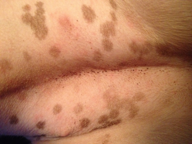
 cutarebra larvae
It sounds like your cat had a Cuterebra Infestation. The Merck Manual describes it better than I can so here is the description:
Etiology:
Adult Cuterebra flies are large and bee-like and do not feed or bite. Females deposit eggs around the openings of animal nests, burrows, along runways of the normal hosts, or on stones or vegetation in these areas. A female fly may deposit 5-15 eggs/site and >2,000 eggs in her lifetime. Animals become infested as they pass through contaminated areas; the eggs hatch in response to heat from a nearby host. In the target host, the larvae enter the body through the mouth or nares during grooming or, less commonly, through open wounds. After penetration, the larvae migrate to various species-specific subcutaneous locations on the body, where they develop and communicate with the air through a breathing pore. After ~30 days, the larvae exit the skin, fall to the soil, and pupate. The duration of the pupation varies depending on the environmental factors and winter diapause.
Clinical Findings and Diagnosis:
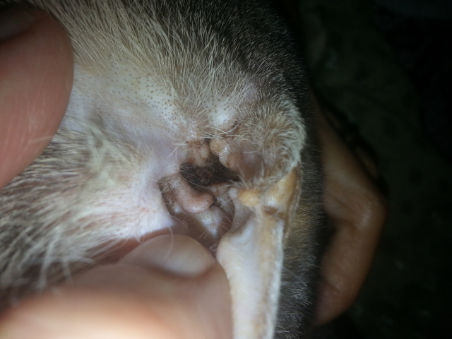
Cuterebra lesions are most common in the summer and fall when the larvae enlarge and produce a fistulous swelling ~1 cm in diameter. Dogs, cats, and ferrets are abnormal hosts for this parasite; aberrant migrations can involve the head, brain, nasal passages, pharynx, and eyelids. In the skin, typical lesions are seen around the head, neck, and trunk. The hair is often matted, and a subcutaneous swelling is present beneath the lesions. Cats often groom the area aggressively. Pain at the site is variable and usually associated with secondary infections. Purulent material may exude from the lesion; the most common differential diagnosis is an abscess or foreign body.
Definitive diagnosis is made by finding and identifying a larva. Second instar larvae are 5-10 mm in length and are gray to cream in color. Third instar larvae are dark, thick, heavily spined and are the stage most commonly seen by veterinarians.
This sounds exactly what your cat had. We have seen this before, so while it is not rare, it isn't uncommon. Once the larvae is out, the wound is healed and that's it. It is not something to worry about, and it will probably never happen again.
There is no sense in asking how this happened, but dealing with it as it is now.
The larvae you saw was probably dead, and that happens quite frequently.
I am glad she is better now. I am enclosing a photo of what the larvae looks like.

 Dull Fur & Flaky Skin
QuestionQUESTION: Can you give your cat the human Omega
Dull Fur & Flaky Skin
QuestionQUESTION: Can you give your cat the human Omega
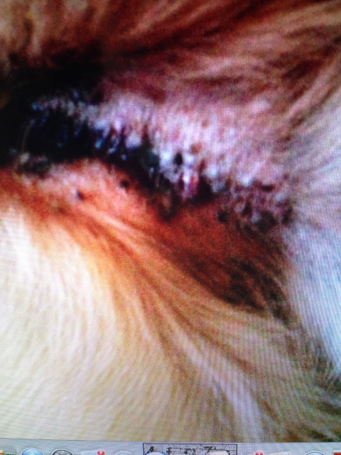
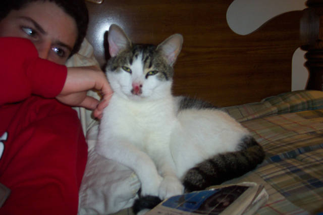 cat with red, raw looking nose, small scabs around it
Question
Raw Nose
My son found a very friendly stray ca
cat with red, raw looking nose, small scabs around it
Question
Raw Nose
My son found a very friendly stray ca
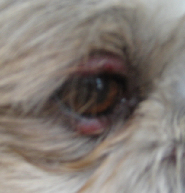 Shih-Tzu eyelide problem
Question
swollen eyelids
Hi,
Its been about 2 weeks no
Shih-Tzu eyelide problem
Question
swollen eyelids
Hi,
Its been about 2 weeks no
 Heartworm treatment side effects
Question
Charlie
My dog, Charlie, is a 30lb. mut
Heartworm treatment side effects
Question
Charlie
My dog, Charlie, is a 30lb. mut