Question
 Ronnie
Hi,
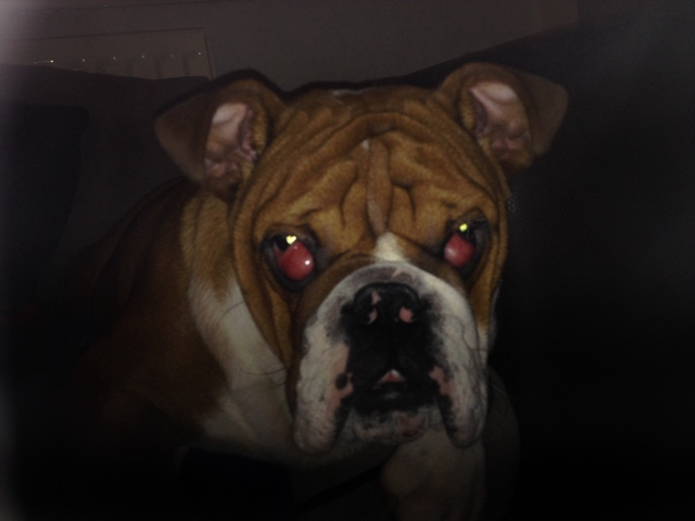
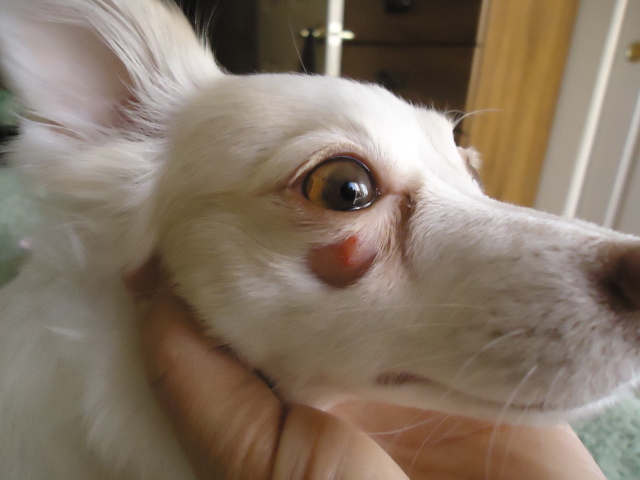
I have a 6 month old english bulldog, who has had cherry eye in both eyes. We took him to the vets and he suggested that we tack his glads back in place with stitches. We where given eye drops and told the swelling would go down. It has now been 8 weeks since is operation and his eyes look worse now, then they did to start with. I have attached a picture for you to look at. Any advise would be grateful as our vet has said that our bulldog will stay like this unless we will need to remove the glands, but he does not advise this, as he could get dry eye.
AnswerHi Dean,
This is a real tough one. I am not a vet so I cannot give you any kind of prognosis with this but I can tell you that while it's true that SOME dogs can get dry eye, it's not that common.
I have seen hundreds of dogs have these removed with no ill effect. This is definitely a breeder issue, however, and you really should advise the breeder of this. It's a congenital defect.
I would ask a second opinion if this were my dog. No dog should have to go through this. Poor guy.
Call a different vet, or call a veterinary ophthalmologist. They specialize in eye care for pets.
Either way, something needs to be done, the sooner the better.
So call around and see a vet you feel comfortable with. Ask friends and family for recommendations as they usually know the good, bad and ugly in vets in your area.
Here is an article on it.Note that they say surgical REPLACEMENT of the gland. That means that they tack in back in there, not remove it. However, it may need to be removed if it doesn't do well."
Cherry Eye:
The enlarged appearance of the third eyelid seen here is due to inflammation of the tear gland.
Cherry eye is the term used for the prolapse of the gland of the third eyelid. It may occur in one or both eyes. The condition is most common in young dogs, six weeks to two years of age. Certain breeds of dogs are predisposed, including the America cocker spaniel, English bulldog, beagle, Chinese shar-pei, Newfoundland, bloodhound, Lhasa apso, miniature poodle, and shih tzu.
Causes
A weakness of the ligamentous attachment of the gland of the third eyelid is believed to be the most common cause in the dog.
Although this weakness may be a heritable condition, the inheritance pattern is unknown.
Prolapse of the gland may occur secondary to inflammation.
Idiopathic (unknown cause) forms also exist.
What to Watch For
Oval pink or red mass protruding from the corner of the eye closest to the nose
Watery or thick discharge from the eye
Redness to the conjunctiva (lining of the eyelid)
Occasional pawing at the eye
Diagnosis
Generally, the diagnosis is made by visual inspection of the eye. A complete eye examination is warranted, including measurement of tear production, fluorescein staining of the cornea and examination of the opposite eye.
Treatment
Medical management involves the use of topical anti-inflammatory corticosteroid medications to decrease inflammation of the conjunctiva and the prolapsed gland. Medical management rarely results in return of the gland to a normal position.
Surgical replacement of the gland is the recommended treatment. Complete removal of the gland may be performed, but it is discouraged because it predisposes the dog to a life of dry eye. The gland of the third eyelid is responsible for the production of around 35 percent of the watery tears, so removal of the gland may result in greatly diminished tear production (dry eye)
Following surgery, an Elizabethan collar may be used to prevent self-induced trauma while the surgery site heals.
Home Care and Prevention
There is a 5 to 20 percent recurrence rate depending on the surgical procedure used, the size of the gland at the time of surgery, the duration of the prolapse, and the condition of the cartilage of the third eyelid. In general, if the gland is replaced quickly, is not too swollen or inflamed, and if the cartilage of the third eyelid is not bent, then the success rate is higher for surgical replacement.
If only one side had prolapsed and was surgically replaced, continue to monitor the other eye for development of a cherry eye. To prevent the other gland from prolapsing, the unaffected gland may be prophylactically sutured at the time the initial gland is operated.
Administer all medication as directed by your veterinarian and return for follow-up as directed by your veterinarian. If the gland stays in place for one month following surgery, then the prognosis is good that it will not reprolapse. If the gland does prolapse again, a second surgical replacement may be attempted, or the gland may be removed.
It is necessary to monitor tear production for sometime after the surgery to determine whether it will remain normal. The onset of dry eye may be delayed for months to years following prolapse of the gland. Signs of dry eye include thick, pussy discharge from the eye, redness to the conjunctiva and cloudiness of the cornea.
It is advisable not to breed dogs that have developed Cherry eye in order to decrease the occurrence of the problem within the breed. "
Some vets can trim the cartilage and not remove the gland itself. You will just have to ask around.
I hope you find one soon. Please let me know how he does.
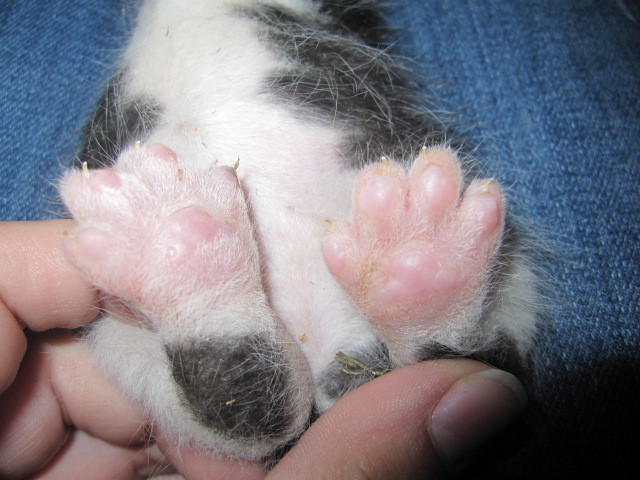 baby kitten w/ all 4 swollen paws
Question
Her back feet
okay... I have 2 young cat
baby kitten w/ all 4 swollen paws
Question
Her back feet
okay... I have 2 young cat
 Worried
Question
eye
My long-hair chihuahua (1.5yr old) has had
Worried
Question
eye
My long-hair chihuahua (1.5yr old) has had
 Runny stool, vomiting,
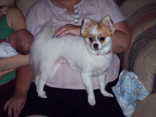
Questionour Pom "Snoball"
QUESTION: Ou
Runny stool, vomiting,
Questionour Pom "Snoball"
QUESTION: Ou
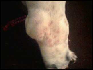 SKIN MISSING PATCHES BUT NO ITCHING
Question
FUR MISSING
I just adopted a pit bull from the
SKIN MISSING PATCHES BUT NO ITCHING
Question
FUR MISSING
I just adopted a pit bull from the
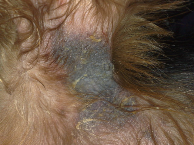 My yorkie has a skin problem
Question
my yorkie
I was wondering if you can help me i
My yorkie has a skin problem
Question
my yorkie
I was wondering if you can help me i