Feeding tubes allow continued feeding of an animal when it has a
mouth, jaw, throat, esophageal, stomach, liver, kidney, or
pancreatic disorder that causes loss of appetite or prevents
normal eating swallowing, or digestion. There are several
different types of feeding tubes, all of which have an external
feeding port connected to a tube that ends in the
gastrointestinal tract.
Nasoesophageal, nasogastric, and nasojejunal tubes enter one of
the nostrils, pass through the nose and throat, and end in the
esophagus, stomach, or small intestine (jejunum), respectively.
Pharyngostomy tubes enter the throat (pharynx) and esophagostomy
tubes enter the esophagus; both end in the esophagus.
Gastrostomy tubes enter the left abdominal wall directly into
the stomach. “PEG” tubes (percutaneous endoscopic gastrostomy
tubes) are gastrostomy tubes that have been placed using
endoscopy. “Button” tubes are short gastrostomy tubes that have
only an external feeding port at the entrance into the body wall
and no external feeding tube. Jejunostomy tubes enter the
abdominal wall and end in the small intestine (jejunum).
Feeding tubes are secured to the body with sutures, bandages,
tube stents, or elastic stockinette or fishnet material. An
Elizabethan collar (or “E-collar”) is sometimes placed around
your pet’s neck to prevent it from removing the tube.
HOW TO FEED (FOLLOW THIS ORDER)
Prepare and warm the food and medications as directed by your
veterinarian. Attach an empty syringe to the external feeding
port. Release or open any external tube clamps (some gastrostomy
tubes have a clamp that should be closed between changing
syringes and between meals). Aspirate the tube with an empty
syringe to check for residual food or fluid left over from the
previous feeding. Return any of the aspirated food or fluid back
into the tube. If more than 20% of the volume of the previous
feeding is aspirated, skip this feeding.
With nasal tubes, administer about 3 mL of water and watch for
signs of coughing or breathing problems; if either of these
occurs, do not give any medication or any food. If liquid
medications are to be given at the time of meal feeding, attach
the medication syringe to the feeding port and administer all
the medications before feeding. Attach a food syringe to the
feeding port and administer the food slowly over 10 to 15
minutes so that your cat can adapt to an enlarging stomach. If
your cat begins to drool or seems uncomfortable, feed more
slowly. (If these signs worsen or your pet vomits during the
feeding, stop feeding.)
Flush the tube with 5 to 10 mL of water (at room or body
temperature). This will help prevent tube clogging. Every time
you finish administering medication or food through the tube,
you must flush the tube with water. Close any tube clamps (if
present). Detach the empty water syringe and close the external
feeding port.
NASOESOPHAGEAL OR NASOGASTRIC TUBES
When feeding, check the tube position and security on the face
and nose. Also check for irritation of the face and nose
(redness, swelling, hair loss, nasal discharge, or sneezing).
Gently remove any debris from the nostril and tube using a warm,
moist cotton ball, gauze, or cloth.
PHARYNGOSTOMY, GASTROSTOMY, AND JEJUNOSTOMY TUBES
Check the tube position daily by locating the external mark
placed on the tube by your veterinarian. Also check the
insertion site for redness, swelling, discharge, or pain. It is
normal for a thin rim of pink or red tissue to grow outward to
the skin of the insertion site. Clean the insertion site with an
antiseptic solution recommended by your veterinarian, and clean
debris on the tube with a warm, moist cotton ball, gauze, or
cloth. After cleaning, place antiseptic ointment and gauze over
the insertion site. You should report the development of
excessive, foul-smelling material to the veterinarian
immediately to circumvent complications from the development of
purulent cellulitisAll of these activities should be performed
daily.
CLOGGED FEEDING TUBES
Check for kinks in the external tube and make sure the tube
clamp is open (if present). Massage the external tube to loosen
any material in the tube. Flush and aspirate the tube with
water. If it flushes but food cannot be administered, check the
tip of the feeding syringe; the syringe tip (versus the tube)
may be obstructed. If water flushing does not relieve the
obstruction, leave water in the tube, and attempt to flush again
in 20 minutes.
WHEN TO CALL THE HOSPITAL
1. The tube position has changed or the tube is no longer secure
or falls out any time. Call immediately.
2. The insertion site or sutured skin is excessively irritated,
swollen, painful, or infected. Call immediately
3. The development of excessive, foul-smelling material. Call
immediately
4. The tube cracks or rips, or its attachments (feeding port,
external stent) become detached. Call immediately
5. Your pet coughs or develops breathing problems. Call
immediately.
6. Your cat vomits, develops a fever, or becomes more lethargic.
Call immediately
7. The tube clogs (even if you can clear it). Your veterinarian
may wish to change the feeding formulation.
FEEDING INSTRUCTIONS
In a mixer, blend _______ (mL or cups) of ______________________
(type of food) with _______ (mL or cups) of water until the
consistency is smooth. Food can be blended and stored in the
refrigerator for up to 3 days. Feed _______ (mL) of the food
mixture slowly over 5 to 10 minutes, _______ times daily. After
each feeding, flush the tube with _______ mL of water.
The above is general veterinary information. Do not begin
any course of treatment without consulting your regular
veterinarian. All animals should be examined at least once every
12 months.
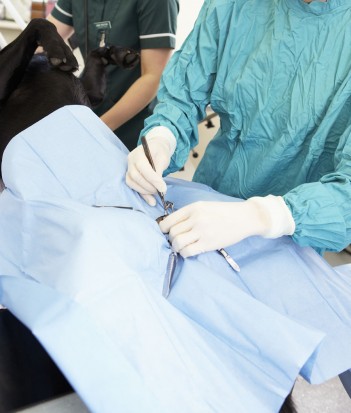 Neutering And Spaying - Is It Morally Right?
Neutering And Spa
Neutering And Spaying - Is It Morally Right?
Neutering And Spa
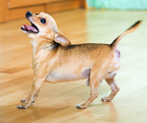 How To Deal With Two Annoying Barking Issues
How To Deal With
How To Deal With Two Annoying Barking Issues
How To Deal With
 Tips for Caring YourPet Better and Keeping Them Happy
Tips for Caring YourPet Better and Keeping Them Happy
Tips for Caring YourPet Better and Keeping Them Happy
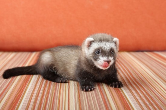 What You Need To Know About Ferret Health
What You Need To
What You Need To Know About Ferret Health
What You Need To
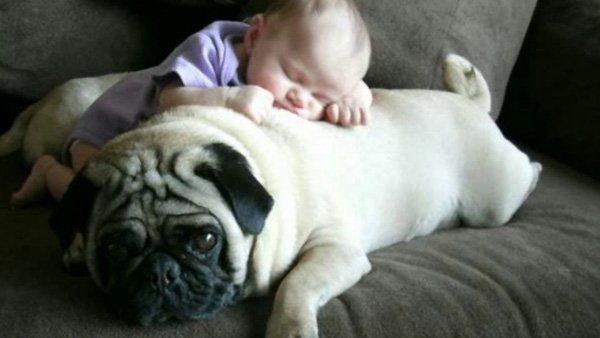
 Should You Let Your Pet Sleep In The Bed?
Should You Let Yo
Should You Let Your Pet Sleep In The Bed?
Should You Let Yo
Copyright © 2005-2016 Pet Information All Rights Reserved
Contact us: www162date@outlook.com