Mast cell tumors are common on or just under the skin of dogs.
Any breed of dog can develop a mast cell tumor (MCT), but
certain breeds are predisposed, including Boxers, bulldogs,
pugs, Boston terriers, golden retrievers, and cocker spaniels.
Mast cells are normal cells within the body that are responsible
for responding to allergic reactions. For example, if you are
slung by a bee and the area becomes red, hot, and itchy, it does
so because mast cells infiltrate into the area, releasing a
variety of substances including histamine, causing these
symptoms. Other than hereditary factors, we do not know why dogs
develop these tumors.
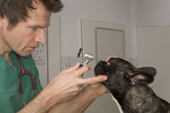
Your veterinarian may have diagnosed this tumor on the basis of
a procedure called fine-needle aspiration. This is a minimally
invasive technique that involves sticking a needle into the
tumor, sucking a few cells out, and smearing the cells on a
slide for a pathologist to evaluate under a micro-scope. This
procedure is not painful to your dog and allows us to make a
diagnosis in most cases. It does not, however, allow us to
predict the biologic behavior of (“prognose”) MCTs; surgical
removal of the tumor followed by the use of a grading system is
required. Location of the MCT is also of prognostic
significance.
Knowing that we are dealing with an MCT before surgery can be
helpful, because MCTs are notorious for sending out long,
finger-like projections of cells into the surrounding tissue.
This means we must surgically remove a wider margin of “normal”
tissue surrounding any visible tumor in an attempt to remove all
the microscopic “fingers.”
Grade I or well-differentiated MCTs are the least aggressive of
the three classes. If we are able to surgically excise the
entire tumor (the pathologist will comment that the margins of
tissue removed are “clean” or free of cancer cells), the
incidence of recurrence is typically small, with 93 per cent of
dogs being disease free at 1 year. “Metastasis” or spread of
this form of MCT to distant, internal locations is unusual.
Grade II or intermediately differentiated MCTs are more
aggressive than their grade I counterparts. An as yet
unidentified percentage of dogs with this form of MCT develop
metastasis of their cancer to intemal organs, typically to the
bone marrow, spleen, or local lymph node. Provided there has
been no spread of the cancer, 50 per cent of dogs with
completely excised grade II or intermediately differentiated
MCTs develop recurrence within 10 months of diagnosis; if no
recurrence is detected in this period of time, there is a very
good chance that the dog will survive for 5 years free of tumor.
Grade III or poorly differentiated MCTs carry a very poor
prognosis, with 97 per cent of dogs succumbing to their cancer
by 1 year. This is due to the high rate of metastasis or spread
of the cancer to internal organs.
Mast cell tumors in the groin behave similarly to grade III
MCTs, regardless of their histologic grade. It is not currently
understood, but a high potential for metastasis has been
consistently observed. Some oncologists believe that MCTs in the
armpits and mucocutaneous junctions (e.g., lip margins, vulva,
anus) can be quite malignant as well.
Once a dog is diagnosed with a MCT, several diagnostic tests are
recommended. First, a complete blood count, bio-chemical
profile, and urinalysis are performed to ensure that your dog
exhibits no negative effects of the cancer in his or her system.
Sometimes, a blood test called a buffy coat test is performed.
This test looks for mast cells circulating through the
bloodstream. This test is useful if it is positive, but it is
often negative even if the cancer has spread; thus, it is not
very sensitive.
The next step is to grade the cancer if this has not yet been
done.
Again, this can be done only by surgically removing all or part
of the tumor. Once the tumor grade is known, a decision
regarding further testing and treatment can be made. If the
local lymph node is enlarged, it will be aspirated to look for
cancer cells. If the MCT has been graded as intermediate or
poorly differentiated (grade II or III), aspiration of the bone
marrow and the spleen is advised. This is the most sensitive
technique for determining whether the cancer has metastasized.
Unfortunately, dogs with mast cell cancer in the bone marrow or
the spleen have a very poor prognosis; many dogs live only 90
days from the time of diagnosis because of the effects of the
cancer cells on the body. Sometimes, even in dogs with advanced
disease, treatment can improve both the quality and quantity of
life. Your veterinarian may refer you to a cancer specialist for
the testing or further discussion of your options for treatment.
Treatment for dogs with MCTs is dependent on the grade of tumor
and results of testing. Dogs with grade I tumors that have been
completely excised (removed) are not typically treated with any
additional therapy. The “gold standard” of treatment for dogs
with grade II MCTs, because of their moderate incidence of local
recurrence even with complete surgical excision, is radiation
therapy.
We also recommend using radiation therapy to treat grade I and
II tumors that cannot be completely excised, provided there is
no evidence of metastasis. Eighty-eight per cent of dogs with
incompletely excised grade II tumors survive for 5 years without
disease when treated with radiation therapy.
For dogs with grade III MCTs, dogs with MCTs in the groin, or
dogs that have been diagnosed with systemic spread of their mast
cell cancer, drug therapy is often recommended. These drugs
include diphenhydramine (Benadryl) and cimetidine (Tagamet) to
counteract the effects of histamine on the body and prednisone
and other chemotherapy drugs to attempt to kill the cancer
cells. These drugs are usually well tolerated by dogs. Signs of
terminal stages of the cancer include lethargy and
gastrointestinal signs such as vomiting, diarrhea, and poor
appetite.
Our goal for all cancer patients is that their quality of life
be excellent; we never want the treatment to be worse than the
disease. This goal is often achieved by working as a close team
with your veterinarian and often a board-certified cancer
specialist.
The above is general veterinary information. Do not begin
any course of treatment without consulting your regular
veterinarian. All animals should be examined at least once every
12 months.

Copyright © 2005-2016 Pet Information All Rights Reserved
Contact us: www162date@outlook.com