Tetanus is an occasional disease in dogs, the result of infection with a bacterium called Clostridium tetani. This bacterium is normally present in soil and other low oxygen environments, but also in the intestines of mammals and in the dead tissue of the wounds that are created due to injury, surgery, burns, frostbite, and fractures.
One typical feature of this bacterium is that it can live without oxygen (anaerobic) and can remain in the environment for long periods by forming spores. Once favorable conditions are present, such as an injured animal coming into contact with the spores, they are able to release the potent toxin into the body. These potent toxins bind to nerve cells in the body and generate symptoms that are characteristic of this disease, such as muscle spasms and stiffening of the limbs.
The severity of the symptoms will often depend on the number of organisms that are able to enter the body and the quantity of toxins produced in the body, but this is generally considered a serious condition warranting immediate treatment.
Symptoms can appear after spores have entered the wound and germinated. The muscles around the infected wound may become rigid first. The dog may appear stiff and lame. Weakness and an uncoordinated gait can usually be observed in these dogs. The symptoms may then disappear spontaneously if the infection remains local to the area in which it entered the body, while in other cases the symptoms can escalate to a generalized disease if the toxins are able to gain access to the nervous system.
The symptoms related to generalized disease are:
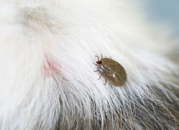
Because unattended wounds leading to bacterial contamination is the leading cause of lockjaw, outdoor dogs are at higher risk.
You will need to give a thorough history of your dog's health, including a background history of symptoms. Your veterinarian will also ask about any previous injuries or traumas that might have led to the infection. After taking a detailed history, your veterinarian will then conduct a complete physical examination on your dog.
Routine laboratory tests will include a complete blood count (CBC), biochemistry profile, and urinalysis. The complete blood count may show an abnormally low or high number of white blood cells (WBCs), both indicating infection. Biochemistry testing may reveal high concentrations of an enzyme called creatine phosphokinase (CPK). This enzyme is mainly found in the heart, brain, and skeletal muscles, but the level of this enzyme increases in the blood in response to the stiffness and damage the muscles are experiencing, which are in turn responding to the bacterial infection.
The results of the urinalysis are often normal except for an increase of myoglobin in the urine. Myoglobin is a protein that is normally found in the muscles, and with constant contractions and stiffness of muscles, it starts appearing in the urine due to its release from the damaged muscles. Your veterinarian will also send samples of tissue and fluid that has been taken from the wound to the laboratory for culture. Culture testing will allow for the controlled growing of the causative organism, thereby confirming its presence in the wound.
In advanced stages of this disease, your dog will need to be hospitalized. Good support and constant nursing is usually required for a period of 3-4 weeks. If your dog is unable to eat on its own, your veterinarian will place a feeding tube directly into its stomach in order to maintain its energy and metabolic needs. Because this toxin attacks the muscles and nervous system, your dog is likely to be very sensitive, making forced feedings an undesirable treatment method. Such manipulations may, in fact, exacerbate the symptoms. Intravenous fluids may be started to prevent dehydration. That will be one of the primary concerns.
One of the important features of nursing care is to keep the dog in an environment of low light and low noise, as these animals are extremely sensitive to touch, sound, and light.
Your dog will be kept sedated to prevent further aggravation of the symptoms. Drugs can be used to minimize the muscle spasm and convulsions. In combination, these types of drugs will encourage your dog to remain in a lying position for extended periods. Because of this, there is a concern for the side effects of lying in one place for too long. You should provide your dog with soft bedding, and you will need to schedule regular times throughout the day when you can turn your dog over to its other side, to prevent bed sores/ulcers from developing.
In the event that your dog is not able to breathe properly, a tube will be placed into the trachea to facilitate normal breathing until the muscles have recovered from the infection. In some animals, a hole has to be made into the trachea to facilitate breathing and prevent asphyxia. If your dog is not able to pass urine, a urinary catheter will be placed to allow for the passage of urine. If your dog is constipated, an enema can be given to relieve constipation. In many cases, these treatments may be applied in the home environment. The most important consideration is the ability to maintain a sterile environment for the dog, if it is going to be receiving home treatment after the initial in-clinic care. You will need to discuss this with your veterinarian and go over the proper procedures for avoiding contamination.
Drugs will be given to bind the toxin and prevent its further binding to the nerve cells. Antibiotics will also also given, either orally or by injection, to control further spread of the infection. Topical (outer) antibiotics will also be used around the periphery of the wound to control infection.
Once your dog is out of danger, you’ll be allowed to take it back home where you will need to provide good nursing care until your dog has fully recovered from the infection and its side effects. Your veterinarian will brief you on the correct usage of the various tubes that will need to be placed in your dog's body, including the stomach tube for daily feeding.
As mentioned above, it is important to change your dog's resting position every few hours to prevent ulcers. Keep the wound clear and visit your veterinarian if you see any change in the color of the wound or if ulcers start appearing. Otherwise, you should expect your dog to feel sore. Your veterinarian will give you pain medication for your dog to help minimize discomfort, and you will need to set up a place in the house where your dog can rest comfortably and quietly, away from other pets, active children, and busy entryways. Trips outdoors for bladder and bowel relief should be kept short and easy for your dog to handle during the recovery period. Use pain medications with caution and follow all directions carefully; one of the most preventable accidents with pets is overdose of medication.
You will need to visit your veterinarian a few more times to have your dog examined and to evaluate its recovery status. The prognosis largely depends on the severity of the disease; the more severe the disease, the less are the chances for a full recovery. Good owner compliance is required as these animals often need a long period of time for a full recovery. A strong commitment from your side will greatly improve your dog's chances for survival.


 Bartonella Infection in Dogs
Canine Bartonellosis
Bartonellosis is an emerging
Bartonella Infection in Dogs
Canine Bartonellosis
Bartonellosis is an emerging
 Abnormal Growths in the Lower Intestines of Dogs
Rectoanal Polyps in Dogs
Rectoanal polyps is char
Abnormal Growths in the Lower Intestines of Dogs
Rectoanal Polyps in Dogs
Rectoanal polyps is char
 Chest Bone Deformity in Dogs
Pectus Excavatum in Dogs
In pectus excavatum, the
Chest Bone Deformity in Dogs
Pectus Excavatum in Dogs
In pectus excavatum, the
 Side Effects of Anxiety Medications in Dogs
Serotonin Syndrome
Dogs suffering from compulsive
Side Effects of Anxiety Medications in Dogs
Serotonin Syndrome
Dogs suffering from compulsive
 Vaginal Inflammation in Dogs
Vaginitis in Dogs
The term vaginitis refers to in
Vaginal Inflammation in Dogs
Vaginitis in Dogs
The term vaginitis refers to in
Copyright © 2005-2016 Pet Information All Rights Reserved
Contact us: www162date@outlook.com