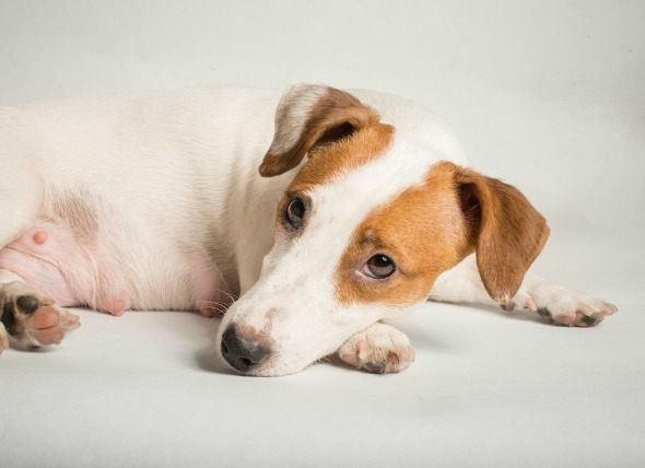
Hepatic nodular hyperplasia is a seemingly benign lesion found in the liver of middle-aged to old dogs. The lesion consists of discrete accumulations of abnormally multiplying (hyperplastic) hepatocytes, the chief functional cells of the liver, and vacuolated hepatocytes – cells that contain fluid or air filled cavities within. This is a cause of high liver enzymes in old dogs.
Clinical findings are derived from associated high liver enzyme activity and ultrasonographic detection of nodules or nodularity in the liver, or by mass lesions observed during exploratory abdominal surgery. Nodular hyperplasia (proliferation of cells) may be mistaken for regeneration secondary to chronic hepatitis, or for a tumor of the liver (adenoma) with needle core biopsies. It may be more common in dogs with vacuolar hepatopathy (liver disease) and may represent a component of that syndrome; it remains uncertain if this is a true syndrome. Although this disease is not breed specific, it may be more common in Scottish terriers. This condition is age-related, with lesions typically developing around six to eight years of age. In one documented clinical study, lesions in all geriatric dogs were found to have occurred in dogs that were greater than 14 years of age.
Nodular hyperplasia does not cause clinical illness unless large nodules rupture and bleed (rare), or the nodules impair hepatic sinusoidal perfusion (delivery of blood to the liver).
An enlarged liver with an irregular hepatic margin (an abnormal border of the liver) may be discovered on a touch exam (palpation), but this is rare. Serendipitous discovery of hepatic nodular hyperplasia during health assessments for other illnesses is common.
Origin is unknown. Metabolic factors that may lead to hepatic nodular hyperplasia are vacuolar hepatopathy (liver disorder), or prior injuries to the liver.
You will need to give a thorough history of your pet's health, including a background history of symptoms, if any, and possible incidents that might have precipitated this condition, such as an injury to the abdominal area. Your veterinarian will perform a complete blood count (CBC), a biochemistry analysis, and a urinalysis. Abdominal radiography, and ultrasonography imaging will allow your doctor to visually examine the liver for abnormalities, along with special stains that can be injected to allow for a visual presentation of the movement of body fluids through the liver organ. A sample of fluid from the liver, taken by aspiration sampling, and a sample of liver tissue by biopsy can also be beneficial in making an accurate diagnosis.
There is usually no treatment required for hepatic nodular hyperplasia. In cases of rupture of large nodules, blood transfusion and emergency mass lesion excision (removal) may be necessary to stabilize your pet.
Your veterinarian will want to perform quarterly biochemical profiles, along with abdominal ultrasonography, to evaluate progression of hepatic nodules, and to head off any subsequent complications that may result from nodular formations impeding the normal functioning of the liver.


 Bleeding Under the Skin of Dogs
Petechia, Ecchymosis, and Bruising in Dogs
Petech
Bleeding Under the Skin of Dogs
Petechia, Ecchymosis, and Bruising in Dogs
Petech
 Anemia Due to Chronic Kidney Disease in Dogs
Erythropoietin (EPO) is a glycoprotein hormone, p
Anemia Due to Chronic Kidney Disease in Dogs
Erythropoietin (EPO) is a glycoprotein hormone, p
 Failure to Absorb Vitamin B12 in Dogs
Cobalamin Malabsorption
Cobalamin malabsorption r
Failure to Absorb Vitamin B12 in Dogs
Cobalamin Malabsorption
Cobalamin malabsorption r
 Black, Tarry Feces due to Presence of Blood in Dogs
Melena in Dogs
The term melena is used to describ
Black, Tarry Feces due to Presence of Blood in Dogs
Melena in Dogs
The term melena is used to describ
 Inflammation of the Soft Tissues in the Mouth in Dogs
Stomatitis in Dogs
Stomatitis is the condition wh
Inflammation of the Soft Tissues in the Mouth in Dogs
Stomatitis in Dogs
Stomatitis is the condition wh
Copyright © 2005-2016 Pet Information All Rights Reserved
Contact us: www162date@outlook.com