Lymph nodes (or glands), are small masses of tissue that can be found throughout the body. They play an integral part in the functioning of the dog's immune system, acting as filters for the blood, and as storage places for white blood cells. Consequently, they are often the first indicators of disease in the tissues.
When tissues become inflamed, the regional lymph nodes that these tissues drain into will also become inflamed and swollen in response. This swelling is due to a reactive increase in white blood cells (hyperplasia) due to the localized presence of an infectious agent. This is medically defined as reactive hyperplasia: when white blood cells and plasma cells (antibody secreting cells) multiply in response to a substance that stimulates their production (antigenic stimulation), causing the lymph node to enlarge.
Lymphadenitis is a condition in which the lymphatic glands have become inflamed due to infection. Neutrophils (the most abundant type of white blood cell, and the first to act against infection), activated macrophages (cells which eat bacteria and other infectious agents), and eosinophils (cells which fight parasites and allergy causing agents) will migrate into the lymph node during an episode of lymphadenitis. This convergence of cells results in the swollen feel and appearance of the nodes.
Cancerous cells may also be found in a lymph node biopsy (tissue sample). Cancer cells may be primary, originating in the lymph node (malignant lymphoma), or may be there as a result of the spread of cancer from another location in the body (metastasis).
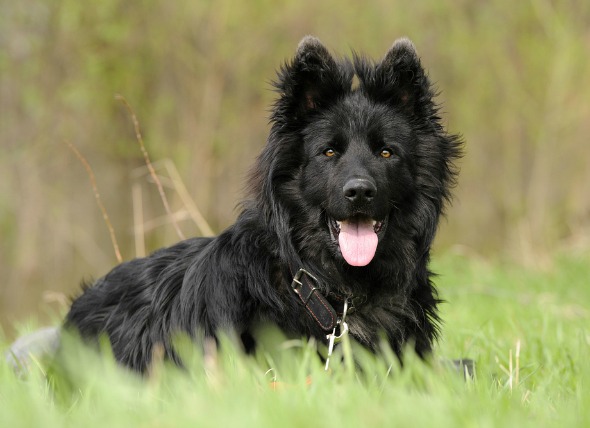
Lymph nodes can usually be detected by touch, but sometimes there will be no clinical symptoms. Swelling can be felt in the area beneath the jaw (submandibular), or around the shoulder. Swelling in one of the legs is also possible as a result of swollen lymph nodes at the back of the leg (popliteal), or near the joint of the leg (axillary – correlating with the armpit). Swollen nodes in the area near the groin (inguinal) may make defecation difficult for your dog. Your dog may also lose its appetite due to nausea, and have an urge to regurgitate when it does eat. You can also expect your dog to feel a general malaise as its body fights off the infection. If your dog has severely enlarged lymph nodes it may have trouble eating, or have difficulty with breathing.
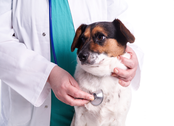
Your veterinarian will perform a thorough physical exam on your dog. A complete blood profile will be conducted, including a chemical blood profile, a complete blood count, an electrolyte panel, urinalysis, and a blood smear.
Lymph node aspirates (liquid) will also be taken for microscopic (cytologic) examination. Abnormal tissue growth, or tumors (neoplasia), and fungal infections can also be confirmed via cytologic examination of lymph node aspirates.
You will need to give a thorough history of your dog's health, including a background history of symptoms, and possible incidents that might have precipitated this condition. The history you provide may give your veterinarian clues as to which organs are causing secondary enlargement of the regional lymph nodes.
Other useful blood tests include serologic (blood serum) tests for antibodies against systemic fungal agents (Blastomyces and Cryptococcus), or bacteria (Bartonella spp.). Radiograph and ultrasound imaging will allow your doctor to visually inspect the affected lymph nodes, and may also enable detection of lesions associated with lymph node enlargement in other organs.
Your veterinarian will prescribe medication dependent on the underlying cause of the lymph node enlargement.
Some infections are zoonotic, meaning that they can be transmitted to humans. Systemic diseases, like sporotrichosis, Francisella tularensis, Yersinia pestis, and Bartonella spp, are zoonotic. If your dog has one of these zoonotic diseases, ask your veterinarian what precautions you will need to take to avoid infection.

 Nerve/Muscle Disorder in Dogs
Myasthenia Gravis in Dogs
Myasthenia gravis is a
Nerve/Muscle Disorder in Dogs
Myasthenia Gravis in Dogs
Myasthenia gravis is a
 Muscle Tear in Dogs
Muscle Rupture in Dogs
A normal muscle can be str
Muscle Tear in Dogs
Muscle Rupture in Dogs
A normal muscle can be str
 Excessive Vocalization in Dogs
Disruptive Crying, Whining and Barking in Dogs
Ex
Excessive Vocalization in Dogs
Disruptive Crying, Whining and Barking in Dogs
Ex
 Toxicity from Gum, Candy, and Toothpaste in Dogs
Xylitol Toxicity in Dogs
There are certain
Toxicity from Gum, Candy, and Toothpaste in Dogs
Xylitol Toxicity in Dogs
There are certain
 High Blood Pressure in the Lungs in Dogs
Pulmonary Hypertension in Dogs
Pulmonary hyperten
High Blood Pressure in the Lungs in Dogs
Pulmonary Hypertension in Dogs
Pulmonary hyperten
Copyright © 2005-2016 Pet Information All Rights Reserved
Contact us: www162date@outlook.com