Pulmonary hypertension occurs when pulmonary arteries/capillaries vasoconstrict (narrow), are obstructed, or receive excessive blood flow. The capillaries of the lungs are very tiny branches of blood vessels only one cell in thickness, connecting the smallest veins to the smallest arteries for the exchange of oxygen and carbon dioxide to the blood and tissues. Arteries carry oxygenated blood from the heart to the lungs, so high blood pressure in the left atrium of the heart can also cause elevated pressure in the capillaries of the lungs.
High pulmonary blood pressure is dangerous because it can alter the shape and performance of the heart. The right ventricle is enlarged, while the left ventricle fills abnormally. Less oxygenated blood reaches the body, leading to troubled breathing, exercise intolerance, and blue-purple tinged skin. Eventually, this increased blood pressure in the right heart can lead to the pooling of blood in the body. The tricuspid valve can also be affected. Located in the right side of the heart, separating the right atrium (upper chamber) from the right ventricle (lower chamber), the tricuspid valve consists of three flaps of tissue that prevent blood from flowing back into the atrium from the ventricle. High pulmonary blood pressure can bring about abnormal functioning of the tricuspid valves, causing a back-flow of blood from the right ventricle back into the right atrium, eventually leading to right-sided congestive heart failure.
Pulmonary hypertension in humans is typically due to an abnormal congenitally formed arrangement of the blood vessels in the lungs (pulmonary vasculature), but with dogs, the current medical findings show that they only develop secondary pulmonary hypertension, that is, hypertension in the lungs due to an underlying disease.
Pulmonary (lung) disease
Extrapulmonary causes of chronic hypoxia (inadequate levels of oxygen reaching the lung tissues)
Your veterinarian will perform a thorough physical exam on your dog, taking into account the background history of symptoms and possible incidents that might have precipitated this condition. You will need to give a thorough history of your dog's health and onset of symptoms. In order to find the underlying cause for the pulmonary disorder, your veterinarian will order a blood chemical profile, a complete blood count, a urinalysis, and an arterial blood gas (ABG) test, to measure the levels of oxygen and carbon dioxide in the blood, as well as to measure the capability of the lungs to move oxygen into the blood. If there is any fluid that has escaped from the vessels into the lining of the lungs (pleura) or abdomen (referred to as effusion), your veterinarian will take a sample for laboratory analysis. If a blood clot in the lung is suspected (pulmonary thrombosis), your veterinarian may perform several more blood tests to confirm this.
A comprehensive examination of the thorax, the cavity where the lungs reside, is essential for diagnosis. Thoracic radiography, or x-ray imaging, is an essential diagnostic tool for your veterinarian to visualize pulmonary abnormalities and/or heart disease. Likewise, an echocardiogram (using Doppler) is a more sensitive tool for finding heart abnormalities, pulmonary blood clots, and measuring pressure gradients in blood vessels when the heart is contracting. Your veterinarian may also use an electrocardiogram (ECG, EKG) to evaluate the electrical functioning of the heart. Recordings from this test would allow your doctor to make a diagnosis based on any observed abnormalities, if they exist, indicating lack of oxygen to the heart muscle.
Your dog will be hospitalized and placed in an oxygen cage if it is showing signs of severe breathing problems. Medicines will be prescribed by your veterinarian in accordance with the underlying disease diagnosis. If the finding is severe heartworm infestation, surgery may be performed to resolve the condition.
Many times the prognosis for secondary pulmonary hypertension is guarded at best. If the disease cannot be resolved, treatment can serve to make your dog more comfortable, but is not curative. If heart failure is diagnosed, your veterinarian will probably prescribe a restricted sodium diet for your dog. Otherwise, to encourage the best conditions for your dog, try to avoid environments that may place undue physical pressure on the dog, such as excessively cold or dry air, excessive heat, second-hand smoke, and high altitudes.


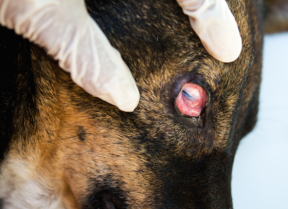 Conjuctivitis (Pink Eye) in Dogs
Conjunctivitis in Dogs
The conjunctiva is the moi
Conjuctivitis (Pink Eye) in Dogs
Conjunctivitis in Dogs
The conjunctiva is the moi
 Addison’s Disease in Dogs
Hypoadrenocorticism in Dogs
Mineralocorticoids an
Addison’s Disease in Dogs
Hypoadrenocorticism in Dogs
Mineralocorticoids an
 Water on the Brain in Dogs
Hydrocephalus in Dogs
Hydrocephalus is an expansi
Water on the Brain in Dogs
Hydrocephalus in Dogs
Hydrocephalus is an expansi
 Hookworms in Dogs
Ancylostomiasis in Dogs
Hookworms can be f
Hookworms in Dogs
Ancylostomiasis in Dogs
Hookworms can be f
 Rapid Heart Beat in Dogs
Supraventricular Tachycardia in Dogs
Supraventric
Rapid Heart Beat in Dogs
Supraventricular Tachycardia in Dogs
Supraventric
Copyright © 2005-2016 Pet Information All Rights Reserved
Contact us: www162date@outlook.com