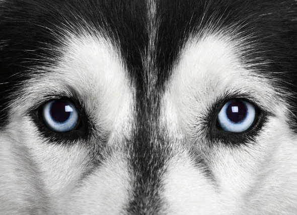
Corneal dystrophy is an inherited progressive condition which affects both eyes, often in the same way. The cornea, the clear outer layer of the front of the eye, is most affected. This disease is not associated with other diseases, and is relatively common in dogs.
There are three types of corneal dystrophy, categorized by location: epithelial corneal dystrophy, where cell formation is affected; stromal corneal dystrophy, where the cornea will become cloudy; and endothelial corneal dystrophy, where the cells of the lining of the cornea are affected.
Epithelial corneal dystrophy:
Stromal corneal dystrophy:
Dog breeds that are predisposed to epithelial and stromal dystrophies:
Endothelial corneal dystrophy:
Dogs breeds that are predisposed to corneal dystrophy:
Epithelial
Stromal
Endothelial
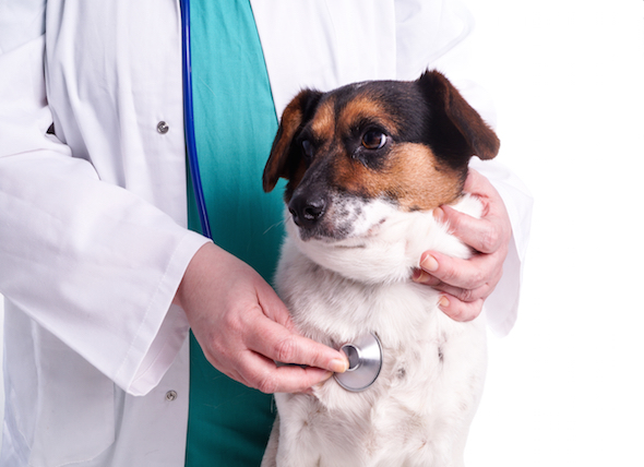
Your veterinarian will perform a thorough physical exam on your dog, including an ophthalmic exam. Your veterinarian will order a blood chemical profile, a complete blood count, an electrolyte panel and a urinalysis. will need to provide a thorough history of your dog's health leading up to the onset of symptoms.
Slit lamp microscopy will aid significantly in differentiating the type of corneal dystrophy present, and a fluorescein stain, a non-invasive dye that shows details of the eye under blue light, will be used to examine the eye for abrasions, and to define the shape of the cornea so that your veterinarian can diagnose the corneal dystrophy. Fluorescein dye enables visualization of any corneal ulcers that may be present; these types of ulcers occur with endothelial and epithelial corneal dystrophy. Fluorescein dye is inconsistent in its ability to aid in diagnosis of endothelial corneal dystrophy, and is not of much use in the diagnosis of stromal corneal dystrophy, but it can be helpful in the diagnosis of epithelial corneal dystrophy. A tonometer will be used to measure the interior pressure in your dog’s eyes so as to rule out glaucoma as a possible cause of corneal swelling.
If your dog has corneal ulcers, they will treated with antibiotic eye medications. Stromal corneal dystrophy usually does not require treatment. Endothelial corneal dystrophy may be treated by using contact lenses over your dog's eyes. Epithelial corneal tags may be removed, if present. Another possible treatment for endothelial corneal dystrophy is flap surgery of the conjunctiva (the lining of the eyeball and the back surface of the lids). While a corneal transplant may be beneficial, results are inconsistent.
Your dog will probably always have some cloudiness to its eyes, even when treatment has been successful. However, if you notice that your dog’s seems to be in pain because of its eyes (e.g., blinking, watering of the eyes) contact your veterinarian, as ulcers may be developing on the cornea. This is prevalent with endothelial and epithelial corneal dystrophy. It is possible that your dog’s vision will remain normal despite corneal dystrophy.
 Inflammation of the Superficial Veins in Dogs
Phlebitis in Dogs
Phlebitis is characterized by a
Inflammation of the Superficial Veins in Dogs
Phlebitis in Dogs
Phlebitis is characterized by a
 Lymph Node Inflammation (Lymphadenopathy) in Dogs
Lymphadenopathy in Dogs
Lymph nodes (or glands),
Lymph Node Inflammation (Lymphadenopathy) in Dogs
Lymphadenopathy in Dogs
Lymph nodes (or glands),
 Dogs and Motion Sickness
Gastrointestinal Distress Related to Motion in Dogs
&nbs
Dogs and Motion Sickness
Gastrointestinal Distress Related to Motion in Dogs
&nbs
 Stomach Worm Infection (Physalopterosis) in Dogs
Physalopterosis in Dogs
Physalopterosis is an inf
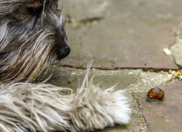
Snail, Slug Bait Poisoning in Dogs
Metaldehyde Poisoning in Dogs
Metaldehyde -- an i
Stomach Worm Infection (Physalopterosis) in Dogs
Physalopterosis in Dogs
Physalopterosis is an inf
Snail, Slug Bait Poisoning in Dogs
Metaldehyde Poisoning in Dogs
Metaldehyde -- an i
Copyright © 2005-2016 Pet Information All Rights Reserved
Contact us: www162date@outlook.com