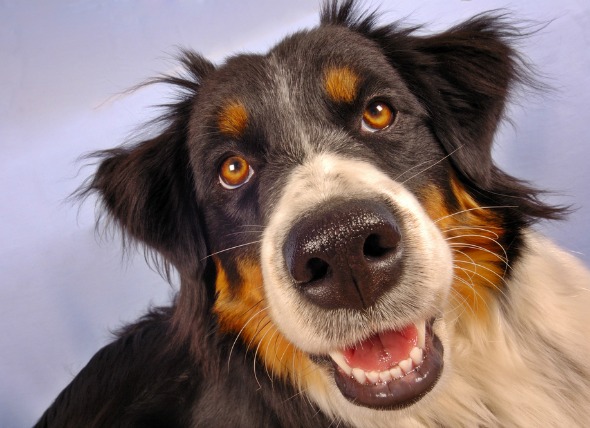
Nonulcerative keratitis is any inflammation of the cornea that does not retain fluorescein stain, a dye that is used to identify ulcers of the cornea. Keratitis is the medical term given to inflammation of the cornea -- the clear outer layer of the front of the eye. If the very top layer of the cornea has been disrupted (as with an ulcer), the dye will enter the lower layers of the cornea and will cause a temporary stain that glows under an ultraviolet light; in nonulcerative keratitis, the top layer of the cornea is not disrupted, so no dye enters the lower layers of the cornea.
In long-term superficial inflammation of the cornea (keratitis), also known as pannus, there may be an inherited susceptibility in the German shepherd and the Belgian Tervuren.
Long-term superficial inflammation of the cornea may occur at any age, but risk is higher between the ages of four to seven years. There are different forms that nonulcerative keratitis can take. Inflammation characterized by the presence of pigment that is deposited in the cornea is sometimes seen in short-nosed, flat-faced (brachycephalic) breeds of dogs, and can occur at any age. In these cases, inflammation of the cornea may be due to exposure to airborne irritants, from a condition in which the eyelids do not close completely, and where there are tear-film deficiencies. Other possible causes include prominent folds of skin around the nose, or abnormal eyelashes that turn inward against the cornea (entropion), which has been notably identified in pugs, Lhasa apsos, shih tzus, and Pekingese.
Inflammation involving the area where the cornea (clear part of the eye) and the sclera (white part of the eye) come together, and characterized by the presence of nodules, may occur in any breed, but is more likely to affect cocker spaniels, greyhounds, collies and Shetland sheepdogs. This form can occur at any age, but varies sometimes by breed. In collies, the average age of occurrence is between three and four years.
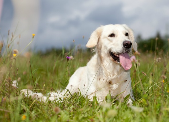
Dry eye is often seen in short-nosed, flat-faced (brachycephalic) breeds, notably cocker spaniels, English bulldogs, Lhasa apsos, shih tzus, pugs, West Highland white terriers, Pekingese, and Cavalier King Charles spaniels. This condition is generally diagnosed in middle to older aged dogs.
Although breed predilection appears to play a role, there is no proven genetic basis in dogs that has been found so far. However, geographical location has been found to play some role, as animals living at higher altitudes appear to be at increased risk.
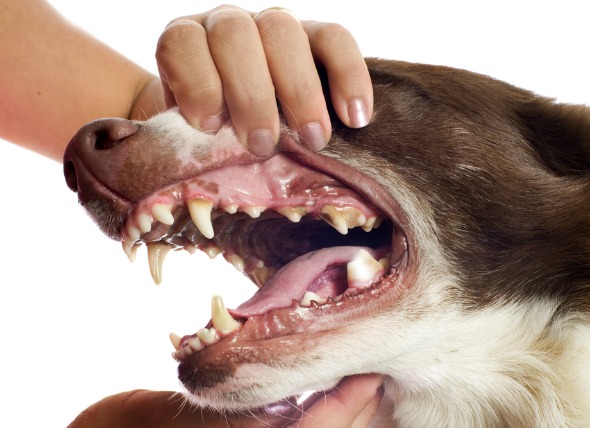
Your veterinarian will perform a thorough physical and ophthalmological exam on your cat, taking into account the background history of symptoms and possible incidents that might have led to this condition. A culture of the fluid in the eye will need to be done. Infectious keratitis is generally easy to diagnose because it is typically ulcerative and painful, distinguishing it from nonulcerative keratitis. If the problem is a tumor, the cornea and sclera are rarely involved. Usually, symptoms will only be on one side. The culture of the cells in the fluid will confirm the diagnosis, and will require a further analysis of the affected eye tissue. A biopsy of the cornea will be done if there are nodules, or if cancer is suspected.
Your dog will only need to be hospitalized if it does not respond adequately to medical therapy. Outpatient care is generally sufficient. Radiation therapy may be prescribed for long-term superficial inflammation of the cornea. Radiation therapy and cryotherapy (a freezing technique that is used for removal of diseased tissue) may also be prescribed for inflammation characterized by the presence of pigment that is deposited in the cornea.
Long-term (chronic) superficial inflammation of the cornea may require surgical removal of the surface of the cornea, but is is only done if the condition is severe; it is usually unnecessary. Even if surgery is performed, indefinite medical treatment will be required to prevent recurrence.
Inflammation that is characterized by the presence of pigment that is deposited in the cornea may also require surgical removal of the surface of the cornea, but it may be performed only after the initial underlying cause is corrected. Surgery is a last resort and is used only in severe cases in which inflammation threatens the dog's vision.
Inflammation that involves the area where the cornea and sclera come together, and that is characterized by the presence of nodules, may require surgical removal of the surface of the cornea. This is usually unnecessary and only temporarily resolves clinical signs; medical treatment will still be required.
If the diagnosis is dry eye, your veterinarian may surgically move the duct from the parotid salivary gland to the eye, in which case the saliva will then compensate for the lack of tears, providing the needed moisture. Surgery may also be necessary to partially close the eyelids.
There are medications that your veterinarian may prescribe as a part of the treatment regimen for the various forms of this condition, and to relieve discomfort.
Long-term superficial inflammation of the cornea in dogs is more likely to occur at high altitudes with intense sunlight.
Your veterinarian will want to conduct periodic eye examinations to evaluate the effectiveness of treatment. Your doctor will set up a follow-up schedule to see your dog at one to two week intervals, gradually lengthening the interval as long as your dog remains in remission, or the clinical signs resolve. In severe cases your dog may have continued eye discomfort, some visual defects, and in some cases, may even suffer from permanent blindness.
 Side Effects of Anxiety Medications in Dogs
Serotonin Syndrome
Dogs suffering from compulsive
Side Effects of Anxiety Medications in Dogs
Serotonin Syndrome
Dogs suffering from compulsive
 Heart Failure, Congestive (Left-sided) in Dogs
Congestive Cardiomyopathy (Left-sided) in Dogs
Th
Heart Failure, Congestive (Left-sided) in Dogs
Congestive Cardiomyopathy (Left-sided) in Dogs
Th
 Acid Reflux in Dogs
Gastroesophageal Reflux in Dogs
Gastroesophageal
Acid Reflux in Dogs
Gastroesophageal Reflux in Dogs
Gastroesophageal
 Dog Diarrhea Treatment And Cures - Diarrhea (Antibiotic-Responsive) in Dogs
Antibiotic-Responsive Diarrhea in Dogs
Veterinari
Dog Diarrhea Treatment And Cures - Diarrhea (Antibiotic-Responsive) in Dogs
Antibiotic-Responsive Diarrhea in Dogs
Veterinari
 Inflammatory Bowel Disease (IBD) in Dogs
The group of gastrointestinal diseases known as i
Inflammatory Bowel Disease (IBD) in Dogs
The group of gastrointestinal diseases known as i
Copyright © 2005-2016 Pet Information All Rights Reserved
Contact us: www162date@outlook.com