When ingested food enters into the intestinal tract, the nutrients and toxins that are a part of the food that has been ingested are released into the digestive blood stream. But before this blood can flow into the systemic blood stream, it first goes through a filtering and detoxification process. The filtering process is carried out mainly by the liver, which detoxifies the blood and sends it out into the main circulatory system. The portal vein, the main part of the hepatic portal system, carries this deoxygenated, prefiltered blood from the digestive tract and its related organs (i.e., the spleen, pancreas and gallbladder) to the liver for processing. When blood pressure in the portal vein reaches a level that is greater than 13 H2O, or 10 mm Hg, this is referred to as portal hypertension. The two main causes of portal hypertension are increased portal flow, or increased resistance to blood.
Increased portal flow occurs when the portal veins attach to arteries, as they do in an arteriovenous fistula (where a new passage is formed between a vein and an artery), or it can occur as a result of blood being diverted (shunted) from the arteries to the liver. Increased resistance to blood can occur in the portal vein prior to its entry into the liver (prehepatic); in the portal vein inside the liver (hepatic); or, it can occur in the hepatic veins in the inferior vena cava (the largest vein in the body, which feeds blood from the lower body to the heart), after blood has exited the liver (posthepatic).
Whether due to increased portal blood flow, or to increased resistance to blood, portal hypertension can cause the formation of multiple portosystemic shunts (PSS), a condition in which the circulatory system bypasses the liver. Animals with portal hypertension can also develop increased abdominal lymph production, leading to fluid build-up in the abdomen. Even more critical is the development of hepatic encephalopathy, which manifests as seizures and problems with mobility due to the unfiltered toxins being delivered to the brain through the bloodstream.
Your veterinarian will perform a thorough physical exam on your dog, which will include a blood chemical profile, a complete blood count and a urinalysis. You will need to give a thorough history of your dog's health leading up to the onset of symptoms.
Other important tests that your doctor will order are tests for total serum bile acids, blood ammonia levels, and sampling of abdominal fluids. Testing the abdominal fluid is essential for determining where the cause of the portal hypertension is originating.
Internal imaging will also be a part of the diagnostic procedures. Results of chest X-rays may show that it is a heart disorder causing the portal hypertension, while abdominal X-rays will allow for a more exact examination of the spleen and liver. An abdominal ultrasound is invaluable for diagnosing disease. In addition, an echocardiogram can assist in diagnosing heart disorders, clots (thrombi), or protrusions in the walls of the abdomen (hernia).
Your veterinarian can also use a diagnostic technique by which the internal anatomy is illuminated by using an injected radioactive tracer. This technique is used for a colorectal scintigraphy, where the colon is examined for abnormalities, and for a portovenography, which allows for examination of the portal system, and which can confirm whether there is a portosystemic shunt (PSS); i.e., a diversion of blood flow. In simple terms, the radiopaque injection (tracer) will allow your doctor to visually examine the blood flow, and see whether the blood is passing through the liver to be filtered and detoxified, or whether the blood is being shunted (diverted) around the liver, creating a toxic condition for the entire system. Angiography, another imaging process using this technique, will allow your doctor to confirm possible abnormal openings and passages (arteriovenous fistulae) in the liver by visually tracing blood flow through the arteries and veins.
A sample of tissue may also need to be taken from the liver (liver biopsy), if liver disease is suspected.
Your dog will probably be hospitalized for monitoring, and for fluid therapy, since dehydration and fluid retention are causes for concern. Your dog's system will need to be detoxed to prevent critical damage to the brain and system.
Surgery may be necessary, but it is dependent on the underlying cause of the disease. If your dog has abdominal fluid build-up, your veterinarian will also prescribe diuretic drugs to treat this.
After your dog has been discharged from care, you will need to restrict its activity until the abdominal swelling has resolved. Dietary changes may be in order, but you will need to consult with your veterinarian before making any major changes to your dog's meals. For example, if your dog has abdominal distention, it may be treated with a low salt diet to moderate the fluid retention, but if your doctor wants to increase urination so that the system will clear, dietary indications will be different, with more fluid intake prescribed.
If your dog is diagnosed with hepatic encephalopathy, your veterinarian will likely recommend a low protein diet, until the liver is at full functioning ability, but again, do not make these changes unless they are recommended by your doctor, since diet changes can have unexpected adverse effects. Your veterinarian will plan follow-up care based on the underlying disease.

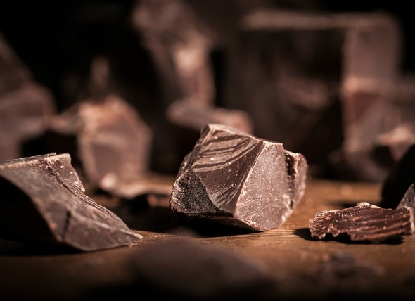 Chocolate Poisoning in Dogs
Dogs are known for eating things when they are not suppo
Chocolate Poisoning in Dogs
Dogs are known for eating things when they are not suppo
 Hematoma on Dogs
Canine Hematoma/Seroma
A hematoma is defined as a
Hematoma on Dogs
Canine Hematoma/Seroma
A hematoma is defined as a
 Liver Failure (Acute) in Dogs
Acute Hepatic Failure in Dogs
Acute hepatic failu
Liver Failure (Acute) in Dogs
Acute Hepatic Failure in Dogs
Acute hepatic failu
 Acne in Dogs
Pustules in Dogs
Much like in teenage humans, acn
Acne in Dogs
Pustules in Dogs
Much like in teenage humans, acn
 Excess Magnesium in the Blood in Dogs
Hypermagnesemia in Dogs
Magnesium is found mostly
Excess Magnesium in the Blood in Dogs
Hypermagnesemia in Dogs
Magnesium is found mostly
Copyright © 2005-2016 Pet Information All Rights Reserved
Contact us: www162date@outlook.com