The stifle joint is the joint between the thigh bone (the femur) and the two lower leg bones (tibia and fibula). It is the quadruped equivalent of the knee in bipeds (i.e., humans).
A ligament is a band of connective or fibrous tissue that connects two bones, or cartilage, at a joint; the cranial cruciate ligament is the ligament that connects the thigh bone with the lower leg bone - it helps to stabilize the stifle joint. Cranial cruciate ligament disease , also referred to as the anterior cruciate ligament (ACL), is the sudden (acute) or progressive failure of the cranial cruciate ligament, which results in partial to complete instability of the stifle joint. Cranial cruciate rupture is the tearing of the cranial cruciate ligament; it is the most common cause of rear-leg lameness in dogs and a major cause of degenerative joint disease (progressive and permanent deterioration of joint cartilage) in the stifle joint; rupture may be partial or complete.
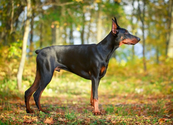
The possibility of a genetic link is unknown. An understanding of the role genetics might play may be important in increasing the likelihood of actively restraining stifle deficiencies and/or structural (conformation) abnormalities. What is currently known is that all breeds are susceptible. Specifically, the incidence of cranial cruciate ligament disease increases for rottweilers and Labrador retrievers younger than four years of age, dogs older than five years of age, and in large-breed dogs from one to two years of age. The predominant gender this affects is the spayed female.
The severity of this condition is related to the degree of rupture: whether it is a partial rupture, or a complete rupture. The manner of rupture is also indicative of the severity, based on whether it presented suddenly, or has been a long-term (chronic) degenerative condition. Degeneration is the decline or loss of function or structure. Sudden (acute) front ligament (cranial cruciate) rupture results in non-weight bearing lameness, and fluid build-up in the joint (known as joint effusion). The dog will hold the affected leg in a partial bent position (flexion) while standing. A subtle to marked intermittent lameness, that may last from weeks to months, is consistent with partial tears in the cruciate; tears that are degenerating and progressing to complete rupture. Normal activity resulting in sudden (acute) lameness would suggest degenerative rupture.
A decrease in muscle mass and weakening of muscles (known as muscle atrophy) in the rear leg - especially the quadriceps muscle group, would be an indication that the leg is not being used properly and the muscles are suffering as a result. Progressive and permanent deterioration of joint cartilage will result if the condition is left untreated, due to ongoing inflammation, and to conditions that will encourage the degeneration of the ligament and surrounding muscles.
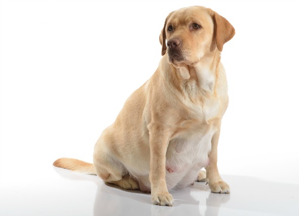
The causes for cranial cruciate ligament disease are most frequently caused by repetitive micro-injury to the cranial cruciate ligament, that is, putting pressure on the ligament in the same way, repeatedly. This action causes slight stretching of the ligament each time, altering the structure, and eventually causing the ligament to tear. Symmetrical or structural abnormalities that occur in the formation, or growth process (conformation abnormalities) are also suspected in the majority of cases. If the bones that make up the stifle were abnormally formed, the cruciate ligament will be unduly stressed and traumatized. Obesity also plays a role in cruciate ligament disease, when it is present, as the weight increases the incidence of repetitious injury to the same part of the leg.
Some of the incidents which may bring about deterioration of the cruciate are injury to the stifle joint; a history of athletics, where repetitive movement can cause stress to the ligaments; a specific traumatic event, as from jumping badly, or any accident that causes the ligament to tear; a knee injury, such as dislocation of the kneecap (medically referred to as patellar luxation).
Your veterinarian will have several diagnostic procedures to follow when looking for the source of the injury. A diagnostic evaluation for cranial cruciate rupture will include a cranial drawer test, which involves specific manipulation to assess the status of the cranial cruciate ligament; puncturing the joint so that fluid can be removed from the point of origin (arthrocentesis), in order to study the cells for toxins, invasions of microorganisms, or immune mediated diseases; and arthroscopy, which uses an arthroscopic tool to directly visualize the interior ligaments, cartilage, and other structures inside and around the joint, as well as to treat abnormalities in the joint.
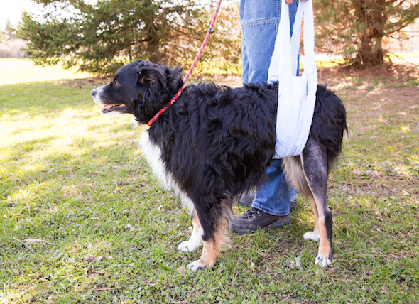
A variety of techniques other than surgery are sometimes used to secure the tibia to the femur and restore stability. An implant can be used to repair the cruciate attachment to the joint. If you wish an alternative to surgery, your veterinarian will be able to advise you on the best course of treatment.
Your veterinarian may also prescribe medications for pain and inflammation if your pet's condition warrants them.
After the condition has been diagnosed and your pet has gone through the initial stage of treatment, management will depend on the particular method of treatment you and your veterinarian decided on. Most surgical techniques require two to four months of rehabilitation. If conformational abnormalities have been determined, it is wise to avoid breeding your pet to prevent passing along the gene. A second surgery may be required in 10 to 15 percent of cases, because of subsequent damage to the meniscus (a crescent-shaped cartilage located between the femur and tibia in the stifle). Regardless of surgical technique, the success rate generally is better than 85 percent.
 Dog Flu
Canine Influenza in Dogs
The virus that ca
Dog Flu
Canine Influenza in Dogs
The virus that ca
 Spasm of the Rear Legs in Dogs
Dancing Doberman Disease
This neurological
Spasm of the Rear Legs in Dogs
Dancing Doberman Disease
This neurological
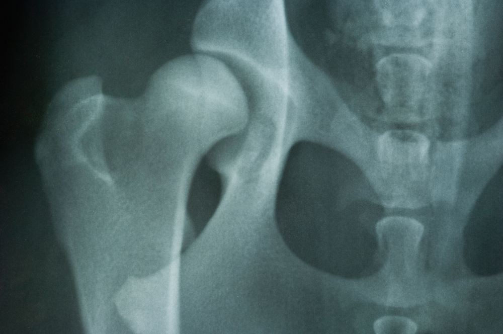 Hip Dysplasia in Dogs
Hip dysplasia in Dogs is a disease of the hip in wh
Hip Dysplasia in Dogs
Hip dysplasia in Dogs is a disease of the hip in wh
 Abortion in Dogs
There are numerous reasons for why pet owners wou
Abortion in Dogs
There are numerous reasons for why pet owners wou
 Nose Cancer (Chondrosarcoma) in Dogs
Chondrosarcoma of the Nasal and Paranasal Sinuses in Dog
Nose Cancer (Chondrosarcoma) in Dogs
Chondrosarcoma of the Nasal and Paranasal Sinuses in Dog
Copyright © 2005-2016 Pet Information All Rights Reserved
Contact us: www162date@outlook.com