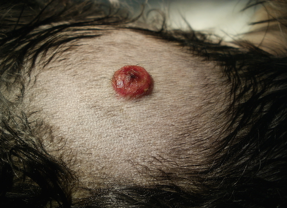
Histiocytic disease are uncommon skin disorders resulting from rapid and excessive growth of cells. This cellular behavior is medically described as cell proliferation.
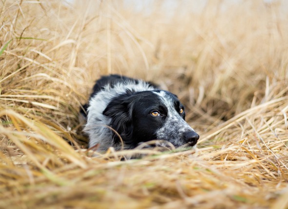
It occurs in young to middle-aged dogs, with a mean age of five years. There is no apparent gender predilection, and skin disease is not restricted to particular breeds, but systemic disease – where the skin disorder spread into the body system – has been reported predominantly in Bernese mountain dogs.
Cutaneous histiocytosis
Malignant histiocytosis
Systemic histiocytosis
Other symptoms and types
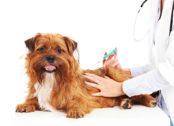
Your veterinarian will perform a thorough physical exam on your dog, including a complete blood profile, chemical blood profile, a complete blood count, and a urinalysis. Extensive laboratory work will need to be essential for making a conclusive diagnosis. A biopsy (tissue and fluid sample) of the affected organs and/or lymph nodes will need to be collected, a cytologic examination (a microscopic examination of the cells) of bone marrow aspiration or biopsy may show systemic histiocytic infiltration. Diagnosis of histiocytosis is often difficult because the results of the microscopic analysis of the cells are not always definitive.
Immunohistochemistry, where a tissue sample is used for the detection of antigens (the molecules that bind to antibodies), typing the tumor and testing the tumor cell's reaction to therapy, can be effectively used for diagnosing a histiocytic disease. Immunohistochemical staining may also be useful for verifying the histiocytic origin of cells.
Fluid therapy or blood transfusions may be required, depending on the clinical findings.
The effectiveness of the treatment is determined by repeated physical examinations, complete blood counts, biochemistry profiles, and diagnostic imaging. The prognosis for dogs with malignant histiocytosis is extremely poor. Death usually occurs within a few months of the diagnosis.
 Liver Inflammation in Dogs
Cholangitis-Cholangiohepatitis Syndrome in Dogs
I
Liver Inflammation in Dogs
Cholangitis-Cholangiohepatitis Syndrome in Dogs
I
 Osteochondritis Dissecans (OCD) in Dogs
Excess Cartilage and Deficient Bone Growth in Dogs
Osteochondritis Dissecans (OCD) in Dogs
Excess Cartilage and Deficient Bone Growth in Dogs
 Distemper in Dogs
Canine Distemper in Dogs
Canine distemper is a co
Distemper in Dogs
Canine Distemper in Dogs
Canine distemper is a co
 Adverse Reaction in Dogs to Glow Stick Jewelry
Dibutyl Phthalate Ingestion in Dogs
Glow sticks a
Adverse Reaction in Dogs to Glow Stick Jewelry
Dibutyl Phthalate Ingestion in Dogs
Glow sticks a
 Runny Nose in Dogs
Nasal Discharge in Dogs
The throat is the end of
Runny Nose in Dogs
Nasal Discharge in Dogs
The throat is the end of
Copyright © 2005-2016 Pet Information All Rights Reserved
Contact us: www162date@outlook.com