In vivo visualization of cardiac function for small animal physiology is essential for the understanding of cardiovascular function in Man. Rat and murine (mouse) models have been developed to investigate cardiac physiology and metabolism in both the normal and diseased heart. Emerging techniques using grasshoppers and zebra fish embryos may enable scientists to understand developmental anomalies in the human heart.
The most common human birth defects are problems with the heart. In utero diagnosis using fetal echocardiography provides the best opportunity to plan and manage these problems once the baby is born. Conditions in the mother where fetal echo may be indicated include diabetes, use of anticonvulsant medication, autoimmune disease, certain viral infections and a previous child with congenital heart disease. Cardiac abnormalities in the fetus detected at 18-week ultrasound scan may also prompt a fetal echo examination.
Fetal echocardiography is safe and accurate. It is capable of detecting almost 100% of all serious heart defects, compared with only a third that are picked up on ultrasound. Challenges associated with fetal echo include the small size of the structures under investigation coupled with random fetal movements. While there are those who argue that fetal echo should be offered to all expectant mothers, others fear that cost and an insufficiency of staff qualified to perform the test make this ideal prospect prohibitive.
Left ventricle (LV) dysfunction is a feature of most cardiac diseases. Speckle-tracking imaging-based regional LV analysis is emerging as a powerful tool for improving the evaluation of LV function. Interference of ultrasound beams within the myocardium (heart muscle tissue) produces characteristic speckle patterns.
Speckle patterns may be tracked using sonomicrometry, a technique that relies on the implantation of piezoelectric transducers in between individual muscle fibers to detect changes in their length. This method allows the detection of minor defects prior to their expression as a global malfunction. Defects may be introduced by genetic engineering or experimental surgical injury models.
Positron emission tomography (PET) scanning produces three-dimensional images of physiological processes in the body. High resolution small animal scanners enable investigations of myocardial blood flow, function and metabolism in the rat heart. This technique is non invasive, thereby permitting the repetitive imaging of the same animal in order to follow disease progression and assessment of new therapies.
Hemolymph is a circulatory fluid found in arthropods and some insects. Synchrotron x-ray phase contrast imaging using microbubbles as high contrast tracer particles has been used to visualize hemolymph flow in the grasshopper. Although the circulatory systems of grasshoppers and humans are vastly different, this technique may be used to elucidate the development of blood flow in vertebrate embryos.
Visualization of cardiac function for small animal physiology in the living organism is critical for our understanding of heart function in our own species. Fetal echocardiography is a safe and accurate means of detecting developing heart problems in humans. Novel techniques such as speckle-tracking imaging, high-resolution PET scanning and synchrotron x-ray phase contrast imaging may one day be used to study normal and diseased heart function in human development.

 Useful Idea For Stronger Nutrition And Wellness
It truly does show when you take time to create a proper no
Useful Idea For Stronger Nutrition And Wellness
It truly does show when you take time to create a proper no
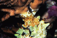 Coral and Invertebrate Quarantine Procedures
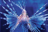 Invasive Aquatic Species
Last column, we discussed invasive species and the potentia
Invasive Aquatic Species
Last column, we discussed invasive species and the potentia
 Aquarium Filtration Done Right
Today, keeping unique aquatic species is now commonplace tha
Aquarium Filtration Done Right
Today, keeping unique aquatic species is now commonplace tha