
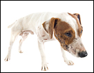
Most pet owners have seen it; your cat or dog hunches up, starts to make retching noises, and then brings up an amount of material. But is it vomit or regurgitation? This may seem like a trivial difference, but in fact it is not. Knowing whether the material is vomit or regurgitation tells your vet where the material is coming from, and also what may be the cause of the problem. Regurgitation is defined as the passive, retrograde movement of ingested material, usually before it reaches the stomach.
The regurgitated material is often expelled with minimal or no signs of nausea, distress or retching. In fact, the animal may show some surprise as it goes through the act, because it was not expecting it! Material originates from the oral cavity, pharynx or esophagus. Regurgitation often occurs immediately after eating food, but can also occur hours afterwards.
If the regurgitation episode happens immediately after eating, there may be something wrong with the upper esophagus. If the material is coated with mucous and lots of saliva, the problem may be with the mid to lower esophagus. Your vet will ask several questions to try and pinpoint the problem. It should also be noted how well the animal eats its food; i.e. can it hold onto the food properly? Is it chewing? Does it look like it is having trouble swallowing? How long have you been observing this problem?
There are three main types of esophageal disorders;
1. Physical obstructions, such as a foreign body stuck in the esophagus.
2) A condition called megaesophagus, where the esophagus is constantly dilated and flaccid.
3) Functional weakness, which may have a nervous cause, such as a damaged nerve.
Obstructions are generally caused by congenital anomalies, foreign objects and the narrowing of the esophagus. The most common congenital reason for obstruction is called a vascular ring anomaly. In this case, the major vessels of the heart are slightly abnormally formed, and actually entrap the esophagus, causing it to be narrowed from the surrounding pressure. A persistent right aortic arch is the most common congenital abnormality. This is correctable by surgery. However, although the episodes of regurgitation may be greatly reduced; they may not be stopped completely. Usually the only signs seen by an owner are young animals (almost always puppies) who are smaller than their littermates, and who regurgitate often. Upon an x-ray or after putting contrast dye into the esophagus, the problem becomes clearer. It is recommended that these puppies never be bred, since this may be a heritable condition.
Megaesophagus is a term used to describe generalized esophageal dilation that is not associated with an obstruction. It may be congenital or acquired. The congenital form is of unknown origin. A predisposition / overrepresentation for congenital megaesophagus has been suggested for the Irish setter, Great Dane, German shepherd, Labrador retriever, Chinese SharPei, Newfoundland, Miniature schnauzer, Fox terrier and also the Siamese cat. Young animals (typically puppies) present with clinical signs of regurgitation, failure to thrive and/or a cough +/- nasal discharge secondary to aspiration pneumonia. As the origin is unknown, conservative medical management is the only known treatment.
In most cases, the cause for megaesophagus is unknown. However, in about a quarter of cases, the underlying cause is a disorder known as Myasthenia Gravis, which is an autoimmune disorder of the nervous system. In either case, supportive treatment such as keeping the animal quiet after eating, and trying to keep the neck elevated for at least ten to thirty minutes after eating is often recommended. This can be done by keeping the dog elevated (i.e.: on its hind legs) if it will allow this.
Finally, a swallowed foreign body can cause acute onset regurgitation, gagging, hypersalivation, discomfort, anorexia and/or odynophagia (painful or exaggerated swallowing). Known or observed ingestion of bones, toys, etc. are an important component of the medical history. Obstruction by smaller objects may go undiagnosed for a longer period of time due to milder clinical signs. Also, if it is a partial obstruction, it may allow for the passage of liquids, and small amounts of food into the stomach while a complete obstruction usually results in regurgitation of both food and water, since almost nothing is actually making it down to the stomach. Secondary signs include coughing, nasal discharge, depression and anorexia. This can happen due to aspiration pneumonia; i.e. the animal has breathed in liquid material into its lungs.
Thoracic radiographs must be done, as well as a barium contrast study if required, to see where the foreign body is lodged. If it does not have sharp edges, it can be removed via endoscopy, or else it may need surgery.
Disorders causing regurgitation can be varied, and this is simply a short summary of some of the causes. Any time your pet repeatedly shows periods of regurgitation, it should be seen by a vet to try and understand the cause and prevent further episodes.
 Puplight Dog Safety Light Red
Using a Puplight is a great
 5 Cute Dog Toy Storage Ideas
If you have a dog and/or mul
 How To Get Rid Of Ticks on Dogs Using Safe and Cheap Alternatives to Frontline Flea and Tick Control
Estimated reading time &ndas
 Dangers of “Free to Good Home” ads
Dangers of “Free to Good Hom
 Choosing the best dog toys for your dog or puppy
Choosing the Best Dog Toys - Plus Our Favorites!
Choosing t
Copyright © 2005-2016 Pet Information All Rights Reserved
Contact us: www162date@outlook.com