Have you ever wondered how cataract surgery is actually performed? If so, read on as it really is a very interesting procedure! Cataract surgery is formally known as phacoemulsification. “Phaco” stands for lens, and to ‘emulsify’ means to break down. This is a great name for this procedure, because this is exactly what it is!
Normally the lens in the eye is a clear soft structure that allows light to easily pass through it. If there is any opacity on the lens, it is known as a cataract. A cataract occurs when proteins in the eye leak into the lens, and start to harden. A general rule of thumb is that if you cannot see into an animal’s eyes clearly, then it can’t see out at you clearly! Thus in a cataract, if the entire lens is opaque due to the protein that has leaked out, then the animal’s vision is reduced.
Cataract surgery involves two main tools; one to break up the cataract into smaller pieces, and the other to vacuum the pieces out of the eye. To begin, the animal is placed under general anesthesia so that it is completely unconscious during the procedure. Then a drug is given to freeze its eye muscles so they absolutely do not move during the surgery. Then the eye is held open with a tiny tool called an eyelid speculum. The area around the eye is carefully draped. The surgery then begins with a small (2-3 mm) long incision made in the eye to introduce the tool that breaks down the cataract. The hard pieces of lens are broken down with ultrasonic waves, and a vacuum tool is introduced to suck it all up. Keep in mind that getting all the pieces out can be difficult; often the lens is stuck in places. It is basically like polishing teeth stained with plaque at the dentist, or trying to get rid of that residue left behind in the bathtub from hard water. Veterinary ophthalmologists who do these surgeries have extremely good hand-eye co-ordination!
After as much of the hard lens is vacuumed out as possible, there is clear view of the eye once again. Then the vet will put in an artificial lens to replace the broken down one; this is called an IOL or IntraOcular Lens. These artificial lenses are NOT cheap, and are specifically designed for animals, and different sizes are available. The tiny lenses are actually foldable, and are pushed into the eye in an amazing way. They are folded into a special tube, the tube is inserted into the empty lens capsule, and then it is pushed into the lens, where it unfolds by itself! Then, a thick, clear solution is pushed into the eye to inflate it, along with a spurt of air.
Finally, the eye is closed up with incredibly thin sutures; so thin that a microscope is needed to work with them. After that, the animal is given pain medication to prevent pain, and then taken off general anesthesia. The aftercare process is truly intense; a huge array of eye drops and ointments are given; some of them have to be given every two hours; around the clock! For this reason, cataract surgeries are often done at veterinary teaching hospitals, where students, residents, interns and vets can all work together to keep treatments on time to prevent complications. Cataract surgery is truly fascinating, and generally 80-90% of animals regain fairly good vision after the procedure!
 Whats in Your Pets Dog Paw and Feet
Paw showing pads A. Claw, B. Digital Pads, C. Metacarpa
Whats in Your Pets Dog Paw and Feet
Paw showing pads A. Claw, B. Digital Pads, C. Metacarpa
 Hypoallergenic Dogs List - The Best Dog Breeds For People With Allergies Or Asthma
Dogs For People With Allergies Or Asthma
Whenever I
Hypoallergenic Dogs List - The Best Dog Breeds For People With Allergies Or Asthma
Dogs For People With Allergies Or Asthma
Whenever I
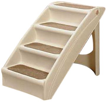
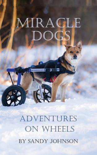 How to Build a Wheelchair for Your Dog
If You Build It, They Will Walk!
How to Build a Wheelchair for Your Dog
If You Build It, They Will Walk!
 German Shepherd Training
Credit: West Midlands Police via Flickr
German Shepherd Training
Credit: West Midlands Police via Flickr
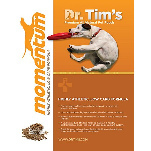
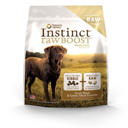
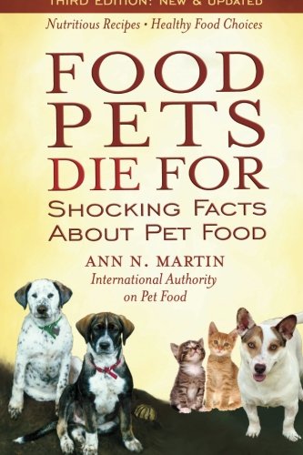 Canine Nutrition and Wellness Part 1: Choosing the Right Commercial Food for Your Dogs
Our canine friends love to e
Canine Nutrition and Wellness Part 1: Choosing the Right Commercial Food for Your Dogs
Our canine friends love to e
Copyright © 2005-2016 Pet Information All Rights Reserved
Contact us: www162date@outlook.com