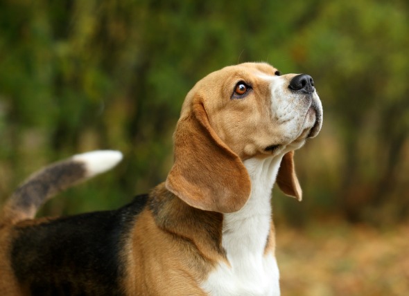
The valves that communicate between the atria and ventricles, the four chambers of the heart, are the atrioventricular valves. The top two chambers of the heart are the atria, and the bottom two chambers of the heart are the ventricles. The mitral valve communicates between the left atrium and left ventricle, and the tricuspid valve communicates between the right atrium and the right ventricle.
Abnormalities in the mitral valve, on the left, affect blood flow to the lungs. The tricuspid valve, on the right side of the heart, is responsible for blood flow to the body. Abnormalities seen here result in poor blood flow throughout the body.
Stenosis (narrowing) of these valves can occur due to the valves being malformed from birth, bacterial heart muscle infection, or cancer of the heart. Stenosis of these valves causes the valves to become leaky, increasing the diastolic pressure gradient between the atrium and the ventricle (the pressure gradient is the period in which the heart's chambers dilate and fill with blood – diastole of the ventricles follows diastole of the atria).
Mitral valve narrowing can cause high blood pressure in the lungs, trouble breathing while exercising, and coughing. Mitral valve stenosis is more commonly seen in Newfoundland and bull terrier breeds.
Tricuspid valve stenosis can lead to swelling of the legs and paws. An enlarged liver may be seen on radiograph images. Tricuspid valve stenosis is more commonly seen in old English sheepdogs and in Labrador retrievers.
Both mitral and tricuspid valve stenosis can lead to congestive heart failure (CHF).
There are a variety of causes that narrow a dog's heart valves, much of it depending on the type of valve stenosis. Mitral valve stenosis, for example, is congenital and commonly affects Bull Terriers and Newfoundlands. Tricuspid valve stenosis, meanwhile, often affects Old English Sheepdogs and Labrador Retrievers, and is also congenital in nature. Moreover, both of these are usually diagnossed at an early age.
Other factors that may lead to narrowing hart valves include cancer of the heart and bacterial infection of the heart muscle.
You will need to give a thorough history of your dog's health and onset of symptoms, including any information you have on your dog's family line. A complete blood profile will be conducted, including a chemical blood profile, a complete blood count, and a urinalysis. The results of these tests typically return normal levels. Based on the apparent symptoms and the results of the initial physical exam, your veterinarian should be able to narrow the cause down to which type of heart valve disease is present. This will need to be confirmed with further testing.
For diagnostic purposes, your veterinarian will need to view the heart using imaging tools. X-rays can help your veterinarian to determine if there is enlargement of the valves or atrium on either side of the heart, and echocardiography will show atrial dilation, and possibly abnormal flow of the blood through the heart. Electrocardiograph readings can also help your doctor to determine if the heart's electrical functioning is being affected. An abnormal rhythm, and the exact measurement of the abnormality can be a great help in determining which side of the heart is most affected.
Your doctor may also use a diagnostic method called angiography, which utilizes x-ray imaging along with a radio-opaque contrasting agent (dye) that is injected into the blood vessels. This dye makes it possible to visualize the vessels internally and evaluate the flow of blood through the heart and surrounding vessels.
In rare instances, a veterinarian may also want to check for pressure disparities within the heart (intracardial) and within the vessels (intravascular) by catheterizing it, a process called cardiac catheterization. This method can also be used for injection of contrasting agents, to take a sample for biopsy, if cancer is suspected, and to assess the severity of the disease.
Medicine is essential to treating heart valve disorders. Diuretics may be used to reduce fluid retention, but other drugs of choice will be based on the final diagnosis. While it is possible to surgically replace or repair damaged valves, it is expensive and has limited availability. An alternative treatment to surgery is a method called balloon valvuloplasty, which may be performed by a specialist after a referral from your veterinarian. Intensive care hospitalization will be necessary for medical treatment of your dog if it is suffering from congestive heart failure.
Your dog will need to be rechecked about every three months or more to see if there are continuing signs of chronic heart failure and to adjust the treatment accordingly. Chest X-rays, an electrocardiogram (EKG – to measure the electrical activity of the heart) and echocardiography will very likely be performed at the follow-up appointments.
Your veterinarian will discuss precautions and home treatment with you, but generally, dogs that are diagnosed with AVD need to be restricted to a low-salt diet and exercise should be restricted.
Because this is a genetically based disease, if your dog is diagnosed with it, your veterinarian will strongly advise against breeding your dog. Spaying or neutering is indicated.
 Kidney Stones in Dogs
Nephrolithiasis in Dogs
Nephrolithiasis is
Kidney Stones in Dogs
Nephrolithiasis in Dogs
Nephrolithiasis is
 Nose Cancer (Adenocarcinoma) in Dogs
Nasal Adenocarcinoma in Dogs
Nose cancer (or nasa
Nose Cancer (Adenocarcinoma) in Dogs
Nasal Adenocarcinoma in Dogs
Nose cancer (or nasa
 Eye Inflammation (Blepharitis) in Dogs
Blepharitis in Dogs
Blepharitis refers to a condi
Eye Inflammation (Blepharitis) in Dogs
Blepharitis in Dogs
Blepharitis refers to a condi
 Tumor of the Thymus in Dogs
Thymoma in Dogs
The thymus is an organ in front o
Tumor of the Thymus in Dogs
Thymoma in Dogs
The thymus is an organ in front o
 Intestinal Disorder (Loss of Motility) in Dogs
Ileus in Dogs
Ileus (functional or paralytic) is
Intestinal Disorder (Loss of Motility) in Dogs
Ileus in Dogs
Ileus (functional or paralytic) is
Copyright © 2005-2016 Pet Information All Rights Reserved
Contact us: www162date@outlook.com