Hemangiosarcomas of the spleen and liver are highly metastatic and malignant vascular neoplasms (tumors in the blood vessels) that arise from the endothelial cells (the cells that line the interior surface of blood vessels). It begins as a large mass that develops in the liver or spleen, spreading rapidly through the blood cell routes, most frequently to the liver from the spleen, or to the lungs from the spleen and liver. In some cases, it can also metastasize to the brain or heart. It can also lead to growth of implantation lesions in the omentum, an apron type fold in the abdominal wall.
Hemangiosarcomas are fed by the blood vessels and fill with blood. Because of this, the tumor can rupture, leading to sudden and severe hemorrhage, collapse, and rapid death. Often, owners do not realize their dog is affected until the sudden hemorrhage or collapse.
In dogs, 0.3 to 2 percent of recorded tumors are found at necropsies; seven percent of all tumors are malignant; and about 50 percent are found in the spleen and five percent in the liver.
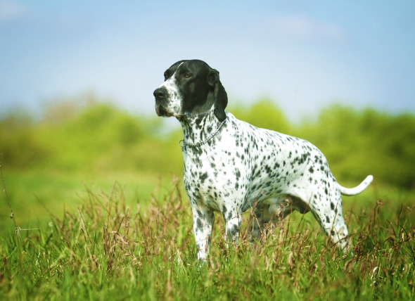
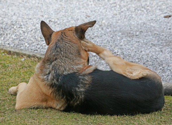
Some dog breeds are more disposed to this type of tumor, including German shepherds, boxers, great Danes, English setters, golden retrievers, and pointers. In addition, there may be a higher risk for male dogs. The average age of occurrence is 8 to 10 years, but it has been seen in dogs younger than one year of age.
Symptoms are generally related to the organs involved; that is, a tumor of the spleen will result in impaired spleen function, and a tumor of the liver will result in impaired liver function. Other common symptoms include:
Cause is unknown.
You will need to give a thorough history of your dog's health leading up to the onset of symptoms, and as much detail as you can about the symptoms you have observed. The history you provide may give your veterinarian clues as to which organs are being affected. A complete blood profile will be conducted, including a chemical blood profile, a complete blood count, and a urinalysis. Findings can include anemia or a low blood platelet count.
Diagnostic imaging is one of the best methods for viewing the abdominal cavity and making an initial diagnosis. X-rays may reveal one or more abdominal masses, along with possible evidence of abdominal fluid. Thoracic radiography of the chest cavity can detect metastasis into the lungs. Ultrasonography can be used to reveals masses in the spleen and any liver involvement. Echocardiography may be performed in patients with evidence of fluid around the heart and may detect cardiac masses. Your doctor may also be able to use ultrasound to guide a fine needle to the tumor in order to take a tissue and fluid biopsy. An analysis of tissue taken directly from the tumor is the most conclusive method for making a diagnosis.
This type of tumor necessitates inpatient care. Intravenous fluids to correct dehydration and transfusions of fresh whole blood for patients with severe anemia will be part of the initial medical care. Coagulation will also be managed as necessary. Depending on the stage of metastasis, surgical management may also be employed. If possible, the tumor will be removed along with the surrounding tissue or the entire organ, A successful splenectomy may give your dog an additional three months of life. If chemotherapy can be successfully employed along with surgery, survival time may be lengthened but not considerably. Because of the aggressive and malignant nature of this tumor, survival time is generally short.
Your dog's activity will need to be restricted until after initial surgical management period is over. Your veterinarian will advise you on the level of activity you should encourage in your dog. It is important to take care in physical activity and to follow your doctor's instructions, since spontaneous hemorrhage may occur.
After surgery, you should expect your dog to feel sore. Your veterinarian may give you pain medication for your dog to help minimize discomfort, and you will need to set up a place in the house where your dog can rest comfortably and quietly, away from other pets, active children, and busy entryways. Trips outdoors for bladder and bowel relief should be kept short and easy for your dog to handle during the recovery period. Use pain medications with caution and follow all directions carefully; one of the most preventable accidents with pets is overdose of medication.
Chest and abdominal radiography and abdominal ultrasound are needed every three months after the initial treatment to monitor for recurrence.


 Degeneration of the Iris in the Eye in Dogs
Iris Atrophy in Dogs
The degeneration of the iris
Degeneration of the Iris in the Eye in Dogs
Iris Atrophy in Dogs
The degeneration of the iris
 Anaerobic Bacterial Infections in Dogs
Anaerobic infections are those that involve bacte
Anaerobic Bacterial Infections in Dogs
Anaerobic infections are those that involve bacte
 Excessive Vocalization in Dogs
Disruptive Crying, Whining and Barking in Dogs
Ex
Excessive Vocalization in Dogs
Disruptive Crying, Whining and Barking in Dogs
Ex
 Skin Mite Dermatitis in Dogs
Cheyletiellosis in Dogs
The Cheyletiella mite is
Skin Mite Dermatitis in Dogs
Cheyletiellosis in Dogs
The Cheyletiella mite is
 'Mad Itch' Pseudorabies Virus Infection in Dogs
Suid Herpesvirus in Dogs
The pseudorabies virus i
'Mad Itch' Pseudorabies Virus Infection in Dogs
Suid Herpesvirus in Dogs
The pseudorabies virus i
Copyright © 2005-2016 Pet Information All Rights Reserved
Contact us: www162date@outlook.com