The thymus is an organ in front of the heart in the rib cage in which T lymphocytes mature and multiply. A thymoma is a tumor originating from the epithelium (layer of tissue covering the thymus) of the thymus. Thymomas are rare tumors in both cats and dogs and they are associated with myasthenia gravis. Myasthenia gravis is a severe autoimmune disease which causes certain muscle groups to tire easily.
Your veterinarian will perform a complete physical exam on the patient. He/she will take a thorough history from the owner. Your veterinarian will order a biochemical profile, a complete blood count, a urinalysis and an electrolyte panel.
Thoracic X-rays should definitely be taken. They may show a cranial mediastinal mass (a mass in between the lungs), pleural effusion (build-up of fluid in the lungs due to aspiration pneumonia) and megaesophagus.
A blood test for antibodies to acetylcholine (a neurotransmitter causing muscles to contract) receptors should be performed so as to rule out myasthenia gravis. A Tensilon test should also be done to test for myasthenia gravis.
A fine-needle aspirate of the mass will show mature lymphocytes (white blood cells) and epithelial cells (cells forming the outside layer of the thymus gland).
Patients should be hospitalized in preparation for surgery to remove the thymoma. They are highly invasive and difficult to remove in dogs. (They are easier to remove in cats.) Dogs with concurrent myasthenia gravis and aspiration pneumonia will have a poorer prognosis despite surgical resection. Twenty to thirty percent of thymomas are malignant and spread throughout the chest and/or abdomen.
If the tumor is completely surgically resectable (and has not spread), the patient will be cured. Your veterinarian will schedule follow-up appointments every three months with you to retake thoracic x-rays of your pet in case the tumor should recur.

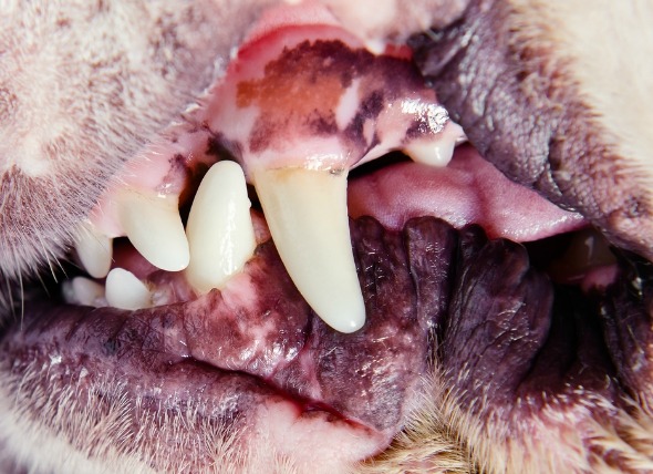 Enlarged Gums in Dogs
Gingival Hyperplasia in Dogs
Gingival hyperplasia
Enlarged Gums in Dogs
Gingival Hyperplasia in Dogs
Gingival hyperplasia
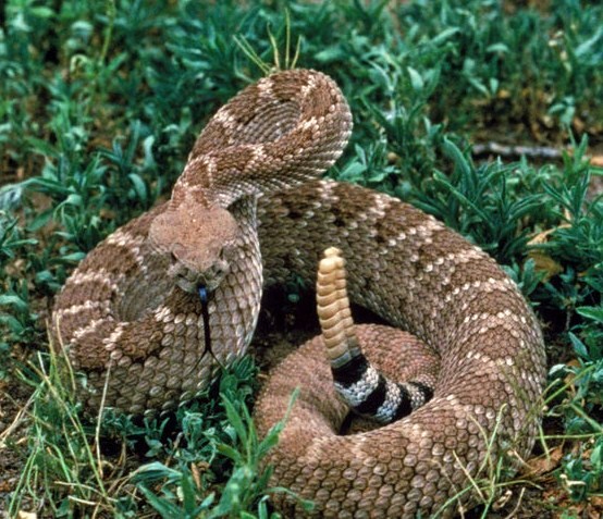 Pit Viper Bite Poisoning in Dogs
Pit Viper Snake Venom Toxicosis in Dogs
Pit viper
Pit Viper Bite Poisoning in Dogs
Pit Viper Snake Venom Toxicosis in Dogs
Pit viper
 Prostate Cancer (Adenocarcinoma) in Dogs
Prostatic Adenocarcinoma in Dogs
The prost
Prostate Cancer (Adenocarcinoma) in Dogs
Prostatic Adenocarcinoma in Dogs
The prost
 Water Mold Infection (Pythiosis) in Dogs
Pythiosis in Dogs
Belonging to the phylum Oomycot
Water Mold Infection (Pythiosis) in Dogs
Pythiosis in Dogs
Belonging to the phylum Oomycot
 Verterbral Disc Inflammation in Dogs
Diskspondylitis in Dogs
Diskspondylitis is the in
Verterbral Disc Inflammation in Dogs
Diskspondylitis in Dogs
Diskspondylitis is the in
Copyright © 2005-2016 Pet Information All Rights Reserved
Contact us: www162date@outlook.com