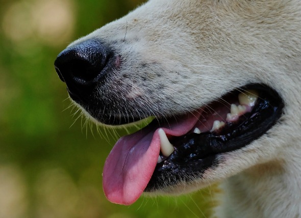
Pemphigus is the general designation for a group of autoimmune skin diseases involving ulceration and crusting of the skin, as well as the formation of fluid-filled sacs and cysts (vesicles), and pus filled lesions (pustules). Some types of pemphigus can also affect the skin tissue of the gums. An autoimmune disease is characterized by the presence of autoantibodies that are produced by the system, but which act against the body's healthy cells and tissues -- just as white blood cells act against infection. In effect, the body is attacking itself. The severity of the disease depends on how deeply the autoantibody deposits into the skin layers. The hallmark sign of pemphigus is a condition called acantholysis, where the skin cells separate and break down because of tissue-bound antibody deposits in the space between cells.
There are four types of pemphigus that affect dogs: pemphigus foliaceus, pemphigus erythematosus, pemphigus vulgaris, and pemphigus vegetans.
In the disease pemphigus foliaceus, the autoantibodies are deposited in the outermost layers of the epidermis, and blisters form on otherwise healthy skin. Pemphigus erythematosus is fairly common, and is a lot like pemphigus foliaceus, but less afflictive. Pemphigus vulgaris, on the other hand, has deeper, and more severe, ulcers because the autoantibody is deposited deep in the skin. Pemphigus vegetans, which affects only dogs, is the rarest form of pemphigus, and seems to be a gentler version of pemphigus vulgaris, with somewhat milder ulcers.
Your veterinarian will perform a thorough physical exam on your dog, including a blood chemical profile, a complete blood count, a urinalysis and an electrolyte panel. Patients with pemphigus will often have normal bloodwork results. You will need to give a thorough history of your dog's health and onset of symptoms. Possible incidents that might have precipitated this condition should also be reported to your veterinarian (e.g., exposure to sun).
A skin exam is crucial. A skin tissue sample will be taken for examination (biopsy); and pustule and crust aspirates (fluid) should be wiped onto a slide to diagnose pemphigus. A positive diagnosis is achieved when acantholytic cells (i.e., separated cells) and neutrophils (white blood cells) are found. A bacterial culture of the skin may be used for identification and treatment of any secondary bacterial infections, and antibiotics will be prescribed in the event that there is a secondary infection present.
Your dog will need to be hospitalized for supportive care if it is severely affected by the condition. Steroid therapy may be prescribed briefly to bring the condition under control. If corticosteroid and azathioprine therapy is prescribed, your dog will be switched to a low-fat diet, since these medications can dispose animals to pancreatitis. Your veterinarian will treat your dog with the drugs that are specifically suited to the form of pemphigus it has.
Your veterinarian will schedule follow-up appointments to see your dog every one to three weeks. Standard blood-work will be performed at each visit to check for progress. Once your dog's condition has gone into remission, it may be seen once every one to three months. The sun can worsen this condition, so it is important to protect your dog from excessive exposure to the sun.
 Bloat or Stomach Dilatation in Dogs
Gastric Dilatation and Volvulus Syndrome in Dogs
Bloat or Stomach Dilatation in Dogs
Gastric Dilatation and Volvulus Syndrome in Dogs
 Irritable Bowel Syndrome in Dogs
Chronic Irritation in the Lining of the Bowels of Dogs
&
Irritable Bowel Syndrome in Dogs
Chronic Irritation in the Lining of the Bowels of Dogs
&
 Nerve Disorder Affecting Multiple Nerves in Dogs
Peripheral Neuropathy (Polyneuropathies) in Dogs
Nerve Disorder Affecting Multiple Nerves in Dogs
Peripheral Neuropathy (Polyneuropathies) in Dogs
 Degeneration of the Image Forming Part of the Eye in Dogs
Retinal Degeneration in Dogs
The retina is the ti
Degeneration of the Image Forming Part of the Eye in Dogs
Retinal Degeneration in Dogs
The retina is the ti
 Swelling of the Salivary Gland in Dogs
Salivary Mucocele in Dogs
An oral or salivary muc
Swelling of the Salivary Gland in Dogs
Salivary Mucocele in Dogs
An oral or salivary muc
Copyright © 2005-2016 Pet Information All Rights Reserved
Contact us: www162date@outlook.com