
Retinal dysplasia in dogs is a condition of the eye, which causes round clumps, sometimes known as “rosettes” or “folds” to form in the retinal tissue. Retinal dysplasia can be caused by a wide variety of factors, but is usually genetically inherited, with various different popular dog breeds being particularly prone to the condition. Retinal dysplasia can affect the vision of the dog in question, but is not painful and is not progressive.
Read on to learn more about retinal dysplasia in dogs, plus find out what breeds and types of dogs are most at risk of developing the condition.
The retina of the eye is composed of two layers, which should form and develop together. Retinal dysplasia occurs when the two layers do not develop properly, causing folds or creases to form between them.
Retinal dysplasia is usually genetically inherited through the breed line and some breeds of dogs in particular are more prone to developing the condition than others (more on this later).
Retinal dysplasia can also be caused by a variety of other means, including vitamin deficiencies, damage to the eye itself, and viral infections that affect the eye. If a bitch is affected by the canine herpes virus during pregnancy, the virus can affect the puppies prior to delivery and potentially cause retinal dysplasia to occur.
There are three different basic types of retinal dysplasia in dogs, which are classed according to their effect on the eye and how they have formed.
Retinal dysplasia in the dog is most usually a hereditary, genetically inherited condition, with some breeds much more prone to presenting with the disorder than others. Breeds within the UK that are most at risk of developing the condition plus the type of the condition that they usually present with are:
With a condition that causes folds or other flaws within the eye, you might reasonably expect that the condition is visible and easy to identify by observation; however, this is not the case. It is not possible to observe the flaws or physical signs of the condition by looking with the naked eye, and so often for dog owners, the first indications that something is amiss is a simple concern that the dog’s vision might not be 100%, for instance if the dog appears particularly clumsy or prone to bumping into things.
In order to definitively identify the condition and rule out any alternative reasons for this behaviour, examination using an otoscope, or even a more complex examination by a specialist veterinary ophthalmologist will be required.
In breeds where the condition is thought to be linked to or associated with a skeletal defect (such as is the case with the Samoyed and the Labrador Retriever) DNA testing can be used to identify the genetic defect that leads to the condition.
Currently there is no way of reversing of treating retinal dysplasia in dogs, and a dog that has been identified to have the condition will have it for life. Dogs that have any form of retinal dysplasia, however mild, should not be used for breeding, as the condition is hereditary.
Depending on how severe the condition is, the associated vision loss that goes with it may be totally manageable, and many pet dogs with retinal dysplasia live full and healthy lives with very little difficulty caused by the condition.
Retinal dysplasia is not progressive, so once the condition is identified it will not worsen over time, although dogs, much like people, may suffer from additional eye problems or failing eyesight as a natural side effect of aging in later life.
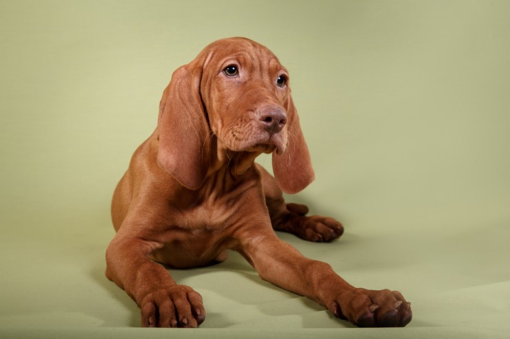 Five Universal Personality Traits Of The Hungarian Vizsla
 Should You Vaccinate Your Dog Against Canine Flu?
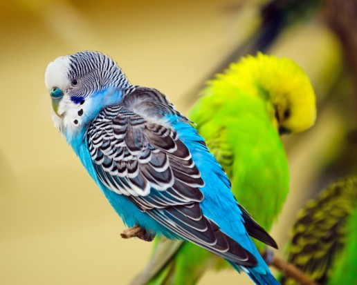 Budgerigar Colour Genetics
 Excellent Chicken Houses from Chicken Coops and Houses for Your Pet
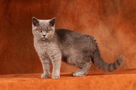 The Feline Calendar – 12 Top Tips To Keep Your Cat Healthy & Happy All Year
 What Is Histoplasmosis Fungal Infection In Dogs ?

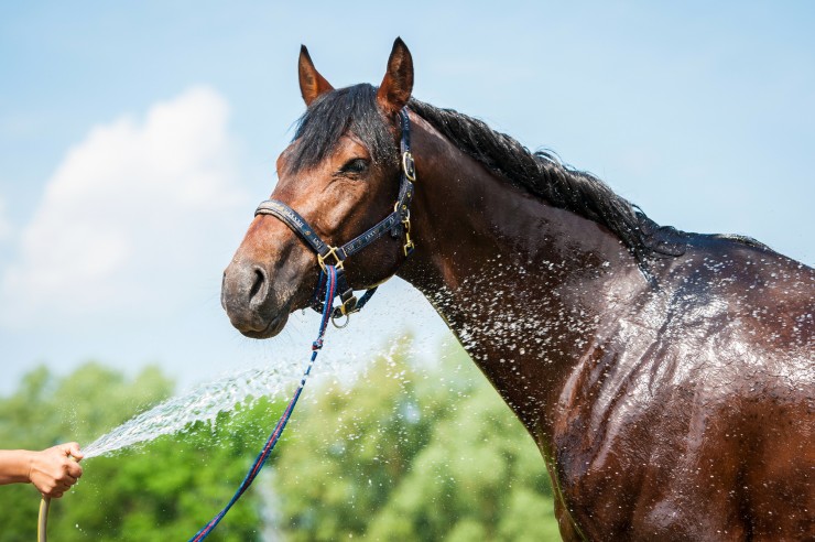 Hot Weather Tips For Horse Owners
Hot Weather Tips
Hot Weather Tips For Horse Owners
Hot Weather Tips
 Keeping Your Cat Off The Kitchen Worktops
Keeping Your Cat
Keeping Your Cat Off The Kitchen Worktops
Keeping Your Cat
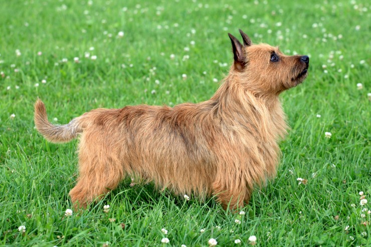 The Australian Terrier As A Family Pet
The Australian Te
The Australian Terrier As A Family Pet
The Australian Te
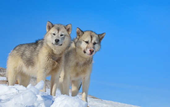 More Information On The Greenland Dog
More Information
More Information On The Greenland Dog
More Information
 English bull terrier puppies – understanding the breed
English bull terrier puppies – understanding the breed
English bull terrier puppies – understanding the breed
Copyright © 2005-2016 Pet Information All Rights Reserved
Contact us: www162date@outlook.com