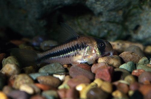
FISH
FISH (fluorescent in SITU hybridization)
FISH (Fluorescent in situ hybridization) is a cytogenetic technique that can be used to detect and localize the presence or absence of specific DNA sequences on chromosomes. It uses fluorescent probes that bind to only those parts of the chromosome with which they show a high degree of sequence similarity
"Fluorescent" means emitting light that comes from a reaction within the emitter. "In situ" refers to the fact that this techniques is done with the chromosomes, cells or tissue in place (in situ) on a microscope slide.
FISH (fluorescent in SITU hybridization)
Multicolor fluorescence in situ hybridization (FISH), in its simplest form, can be used to identify as many labeled features as there are different fluorophores used in the hybridization. By using not only single colors, but also combinations of colors, many more labeled features can be simultaneously detected in individual cells using digital imaging microscopy.
DNA is a double stranded molecule, and when it is chemically denatured and separated into two strands, it quickly reanneals into a double stranded conformation. Thus, when a single stranded probe is incubated with a single-stranded (denatured) metaphase chromosome (or interphase), the probe will bind to complementary DNA sequences to reform the double stranded molecule. Overall, the most critical step when using FISH is the choice of adequate probes. A DNA probe is defined according to its target or complementary DNA in metaphase and interphase cells: (1) repetitive sequence probe, (2) whole chromosome (painting), (3) locus-specific probes.
Repetitive sequence probe are generated from repetitive sequences found in the middle of each chromosome. Researchers use these probes to determine whether an individual has the correct number of chromosomes. These probes can also be used in combination with "locus specific probes" to determine whether an individual is missing genetic material from a particular chromosome.
FISH (fluorescent in SITU hybridization)
Whole chromosome probes are actually collections of smaller probes, each of which binds to a different sequence along the length of a given chromosome. Using multiple probes labeled with a mixture of different fluorescent dyes, scientists are able to label each chromosome in its own unique color. The resulting full-color map of the chromosome is known as a spectral karyotype. Whole chromosome probes are particularly useful for examining chromosomal abnormalities, for example, when a piece of one chromosome is attached to the end of another chromosome.
Locus-specific probes are bind to a particular region of a chromosome. This type of probe is useful when scientists have isolated a small portion of a gene and want to determine on which chromosome the gene is located.
DNA is a double stranded molecule, and when it is chemically denatured and separated into two strands, it quickly reanneals into a double stranded conformation. Thus, when a single stranded probe is incubated with a single-stranded (denatured) metaphase chromosome (or interphase), the probe will bind to complementary DNA sequences to reform the double stranded molecule. Overall, the most critical step when using FISH is the choice of adequate probes. A DNA probe is defined according to its target or complementary DNA in metaphase and interphase cells: (1) repetitive sequence probe, (2) whole chromosome (painting), (3) locus-specific probes.
Repetitive sequence probe are generated from repetitive sequences found in the middle of each chromosome. Researchers use these probes to determine whether an individual has the correct number of chromosomes. These probes can also be used in combination with "locus specific probes" to determine whether an individual is missing genetic material from a particular chromosome.
FISH (fluorescent in SITU hybridization)
Whole chromosome probes are actually collections of smaller probes, each of which binds to a different sequence along the length of a given chromosome. Using multiple probes labeled with a mixture of different fluorescent dyes, scientists are able to label each chromosome in its own unique color. The resulting full-color map of the chromosome is known as a spectral karyotype. Whole chromosome probes are particularly useful for examining chromosomal abnormalities, for example, when a piece of one chromosome is attached to the end of another chromosome.
Please log on to : http://www.ivfsurrogacy.com
Please log on to : http://www.ivfsurrogacy.com/genetic/Fish_fluoroscent_insitu_hybridization.htm
contact Email : info@wecareindia.com
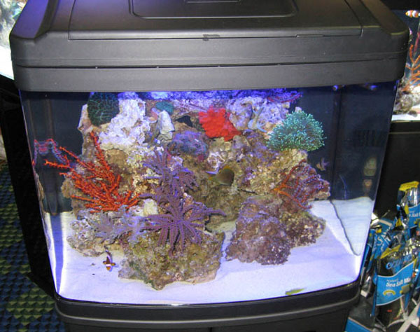
 Red Algae in Marine Aquarium
Q. Ive been keeping saltwater fish for about five years, and
Red Algae in Marine Aquarium
Q. Ive been keeping saltwater fish for about five years, and
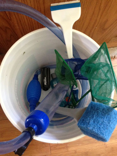 What to Keep in Your Aquarium Maintenance Bucket
It is too easy for hobbyists, especially new ones, to become
What to Keep in Your Aquarium Maintenance Bucket
It is too easy for hobbyists, especially new ones, to become
 How to Get Rid of Cloudy Aquarium Water
Occasional cloudy water is an issue for nearly all aquarists
How to Get Rid of Cloudy Aquarium Water
Occasional cloudy water is an issue for nearly all aquarists
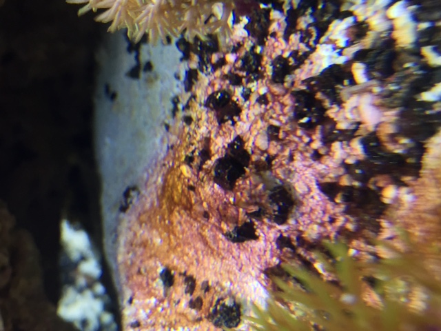
 Aiptasia
Q. I have Aiptasia all over my aquarium and have been
Aiptasia
Q. I have Aiptasia all over my aquarium and have been