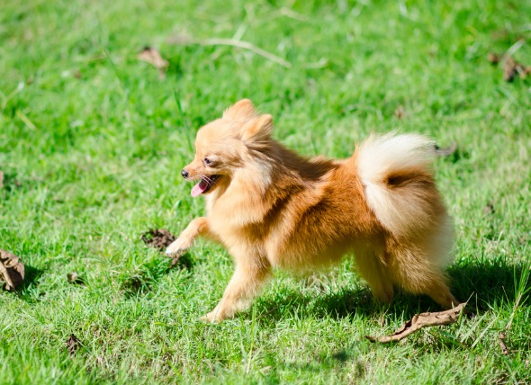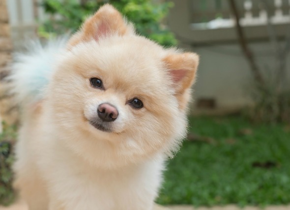
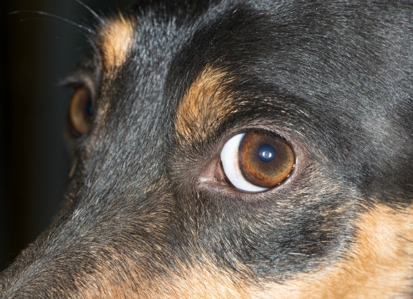
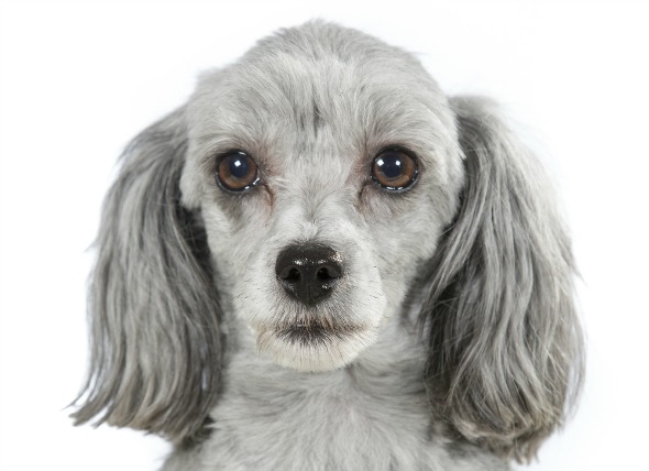
Epiphora is a condition that causes an abnormal overflow of tears. Causes of epiphora due to the shape of the eyes is seen in many breeds. The overproduction of tears can be congenital due to distichiasis – turning in of the eyelashes, or entropion – the turning in of the eyelid. Young shelties, shih tzus, Lhasa apsos, cocker spaniels, pekingese, bulldogs, dachshunds, and miniature poodles are most commonly affected with distichia. Entropion is most commonly seen in some Chinese shar peis, pugs, mastiffs, poodles, Labrador retrievers and chow chows. The upper or lower lid may be affected. This condition may occur secondary to eye irritation.
Epiphora is evident with the observation of an overflow of tears, tear drainage and/or staining on face. Other signs include:
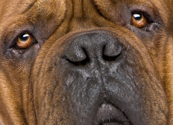
Congenital abnormalities include the occurrence of too large an opening of the eyelids, causing increased exposure of the eyeball in brachycephalic breeds. Ectropion, a turning outward of the eyelid, is commonly found in Great Danes, bloodhounds, and spaniels. Entropion is seen at birth in some breeds and can be acquired due to post-traumatic eyelid scarring and facial nerve paralysis.
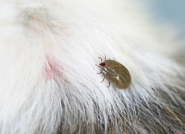
Conditions acquired by a dog can lead to epiphora. These conditions include rhinitis/sinusitis, which causes swelling adjacent to the tear drainage system; trauma or fractures of the bones in the face; foreign bodies in the eyes (e.g., grass, seeds, sand, parasites). Tumors of the third eyelid, the conjunctiva of the eye, eyelids, nasal cavity, maxillary bone in the face, or in the sinuses located around the eyes will also be considered. A condition which causes the nasolacrimal duct (tear duct) to be obstructed, whether through inflammation due to an acquired condition, or because of a congenital abnormality, may also cause an overflow of tears.
Blockage of the nasolacrimal drainage system can be caused by congenital lack of normal openings on the eyelids into the tear drainage system, as seen in cocker spaniels, bulldogs, and poodles. Extra openings can also form into the tear drainage system in abnormal positions, such as openings along the side of the face below the corner of the eye, closest to the nose. Other possibilities include lack of openings from the tear drainage system into the nose.
Acquired conditions involving corneal or conjunctival foreign bodies are seen usually in young, active, large-breed dogs. Inflammation of the eyelids and conjunctiva can be due to infectious or immune-mediated causes. Disorders of the cornea are characterized by the presence of scratches/ulcers with or without inflammation. Inflammation of the front part of the eye, including the iris, can be present. Glaucoma is a condition in which the pressure within the eye is increased. Eyelid tumors are typically seen in older dogs of all breeds.
Your veterinarian will perform a thorough physical exam on your pet, taking into account the background history of symptoms and possible incidents that might have precipitated this condition.
Your veterinarian may order radiographs to check for lesions in the nose or sinus area, and contrast material may be used to help differentiate structures. Your doctor may also order a magnetic resonance imaging (MRI) or computed tomography (CT) scan. In addition, a culture of the material in the eyes will be taken for laboratory analysis. However, surgical exploration may be the only way to obtain a definitive diagnosis. A flushing of the tear ducts may be sued to dislodge any foreign material.
If irritation is evident, your veterinarian may also employ the use of a fluorescein stain, a non-invasive dye that shows details of the eye under blue light, in order to examine the eye for abrasions or foreign objects.
The first step in treatment will be to resolve the cause of the eye irritation ‒ i.e., remove the foreign body from the moist tissues of the eye or the cornea/sclera. Treatment of the primary eye disease, such as conjunctivitis, corneal ulceration with or without inflammation, and/or inflammation of the iris and other areas in the front part of the eye will be the priority. Successful management of a primary lesion that is blocking drainage of tears may allow normal tear flow through the tear drainage system to resume. Patients with inflammation of the nasolacrimal sac may need a catheter placed in the tear duct to hold it open and to prevent scar formation
If the cause is abnormal eyelid formation, surgical repair may be necessary. This is typically a straightforward procedure, where the lids are tacked into a normal position and allowed to readjust. Healing is normally quick and the condition is satisfactorily resolved.
Cryosurgery or removal of hair by electrolysis can be used to treat distichiasis.
Your veterinarian will prescribe appropriate medications based on the diagnosis and plan for treating your dog. These may include topical antibiotic ointments and pain relieving ointments that will contribute to the healing process. An Elizabethan collar should be used during the time of recovery to prevent your dog from further irritating the site.
If your dog has been suffering from inflammation of the nasolacrimal sac, your veterinarian will want to reevaluate your dog every seven days until the condition has been resolved. Treatment will be continued for at least seven days after resolution to help prevent recurrence. If the problem persists for more than 7-10 days, with treatment, or recurs soon after cessation of treatment, a foreign body or persistent infection may be involved, and your veterinarian will want to carry the diagnostic efforts further.
If a surgical procedure to create an opening to drain tears into the nasal cavity was performed, the tubing, called a canula, will be reevaluated every seven days to ensure that it has remained intact. The canula may need to be resutured in place if it becomes loosened or dislodged. After the tubing has been removed, it will be reevaluated again in 14 days.
Recurrence is the most common complication of this condition. This is usually caused by a recurrence of the cause of the eye irritation; a recurrence of inflammation of the nasolacrimal sac; or closure of the surgical openings that were created to allow tears to drain into the nasal cavity
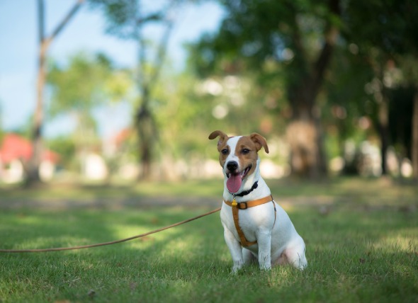
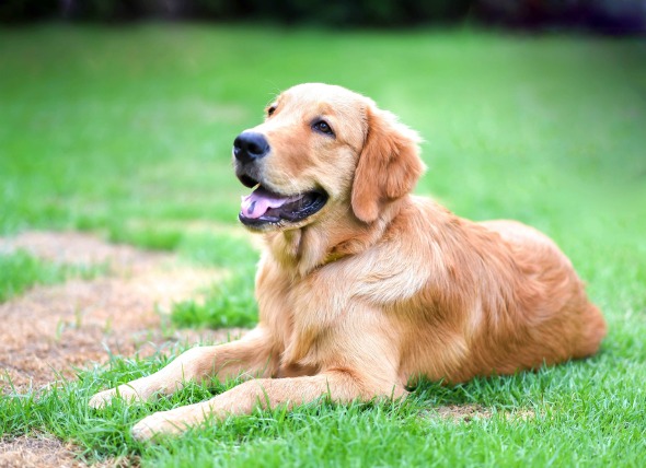
Active outdoor dogs are at risk for being affected by foreign bodies in the eyes.
 Cancer of the Blood Vessel Cells in Dogs
Hemangiopericytoma in Dogs
A hemangiopericytoma i
Cancer of the Blood Vessel Cells in Dogs
Hemangiopericytoma in Dogs
A hemangiopericytoma i
 Abscesses in Dogs
Surface wounds are fairly common in pets, but the
Abscesses in Dogs
Surface wounds are fairly common in pets, but the
 Gallbladder and Bile Duct Inflammation in Dogs
Cholecystitis and Choledochitis in Dogs
The gallb
Gallbladder and Bile Duct Inflammation in Dogs
Cholecystitis and Choledochitis in Dogs
The gallb
 Glaucoma in Dogs
Disease of the Optic Nerve in Dogs
Glaucoma is a
Glaucoma in Dogs
Disease of the Optic Nerve in Dogs
Glaucoma is a
 Skin Rash Due to Contact with Irritants in Dogs
Contact Dermatitis in Dogs
Contact dermatitis may
Skin Rash Due to Contact with Irritants in Dogs
Contact Dermatitis in Dogs
Contact dermatitis may
Copyright © 2005-2016 Pet Information All Rights Reserved
Contact us: www162date@outlook.com