
Infiltrative lipoma is a variant tumor that does not metastasize (spread), but which is known to infiltrate the soft tissues, notably the muscles. It is an invasive, benign tumor composed of fatty tissue, and while it is known mainly for its penetration into muscular tissue, it is also commonly found in the fasciae (the soft tissue component of the connective tissue system), tendons, nerves, blood vessels, salivary glands, lymph nodes, joint capsules, and occasionally the bones. Muscle infiltration is often so extensive that surgery cannot be performed without severe consequences.
Infiltrative lipoma occurs much less frequently than does lipoma. When it does occur, it is usually in middle-aged dogs, and it tends to affect females more so than males. Labrador retrievers are suspected to be at higher risk.
You will need to give a thorough history of your dog's health and onset of symptoms. Your veterinarian will use X-ray imaging to reveal the fat dense tissue between the soft tissue dense structures, and a computed tomography (CT) scan will help to discriminate the nature of the tumor so that your doctor can plan what type of radiation treatment would be best. However, differentiating normal fat from an infiltrative lipoma can be very complicated and problematic.
A sample of tumor cells may be taken by needle aspirate for laboratory analysis, and this may help your doctor to distinguish between normal adipose (fatty) tissue and a lipoma tumor. Lipoma tumors do have a distinctive feature in that they infiltrate muscles, so your doctor may be able to make a form diagnosis based on their behavior within the muscular structure.
The characteristic deep invasiveness of this tumor, along with the difficulty in distinguishing between the tumor and normal fatty tissue, makes removal extremely difficult. Poorly defined tumor margins, the edges of the tumor mass, may also contribute to the high recurrence rate after surgical excision has been performed. A high percentage of post operative patients suffer recurrence within 3–16 months, at a rate estimated at 36–50 percent.
There is an exception, and that is when a tumor has been located in one of the limbs and the entire limb removed. However, amputation of an affected limb is recommended only when quality of life is affected, since these tumors cause little inconvenience unless they interfere with movement, cause pressure-related pain, or develop in a vitally important site, such as a major blood vessel. Amputation is also recommended before growth of the tumor can cross an attainable surgical margin.
Radiotherapy can be beneficial for long-term tumor control. A median survival rate of 40 months was estimated in a retrospective study of 13 dogs, with only one dog euthanized. Dogs with measurable disease may only have stabilization of the tumor (meaning, no further disruption of health. Your veterinarian will prescribe only those medications that have a direct relationship to the treatment method, such as those that will stop or slow tissue growth.


 Nose and Sinus cancer (Squamous Cell Carcinoma) in Dogs
Nasal Squamous Cell Carcinoma in Dogs
The respira
Nose and Sinus cancer (Squamous Cell Carcinoma) in Dogs
Nasal Squamous Cell Carcinoma in Dogs
The respira
 Heart Defect (Congenital) in Dogs
Patent Ductus Arteriosus in Dogs
The aorta is the
Heart Defect (Congenital) in Dogs
Patent Ductus Arteriosus in Dogs
The aorta is the
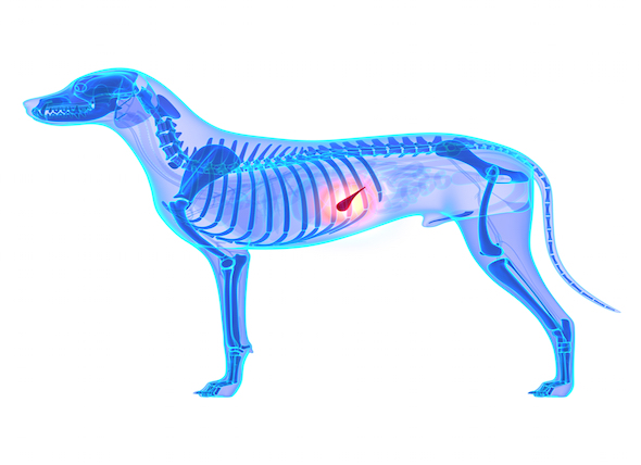 Inflammation of the Pancreas in Dogs
Pancreatitis in Dogs
The pancreas is part of the
Inflammation of the Pancreas in Dogs
Pancreatitis in Dogs
The pancreas is part of the
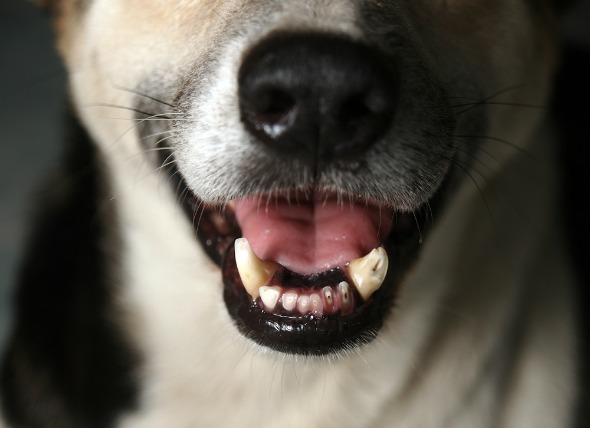
 Teeth Misalignment in Dogs
Malocclusion of Teeth in Dogs
Normally, a p
Teeth Misalignment in Dogs
Malocclusion of Teeth in Dogs
Normally, a p
 Lung Cancer (Adenocarcinoma) in Dogs
Adenocarcinoma of the Lung in Dogs
Adenoca
Lung Cancer (Adenocarcinoma) in Dogs
Adenocarcinoma of the Lung in Dogs
Adenoca
Copyright © 2005-2016 Pet Information All Rights Reserved
Contact us: www162date@outlook.com