Hyphema, or blood in the anterior chamber of the eye, is a common condition among dogs. However, hyphema is a clinical sign and not a specific disease.
The symptoms of hyphema are dependent on the extent of bleeding, whether vision has been impaired, and whether your dog has other, underlying systemic diseases.
Common signs that are observed during a physical examination are:
The most common causes of hyphema are:
Hyphema can also be indicative of various ocular (eye) and systemic deficiencies, some of which may be life threatening. Therefore, its diagnosis and proper treatment is very important.
Hyphema is diagnosed through hematology and blood biochemistry tests, lab tests, and diagnostic imaging using X-rays and ultrasound tests.
A complete medical history will be taken and a thorough physical examination done to include or exclude possible causes for the condition.
Common diagnostic tests and procedures include:
Other advanced tests that may be performed include abdominal ultrasounds, X-rays of the head and eye orbit to detect hitherto unknown traumatic injuries, and hormonal tests (assays) of the adrenal glands. To detect bone marrow cancer, a bone marrow aspirate - the liquid found within the bone marrow - may also be done.
The objectives of hyphema treatment involve containing the inflammation and removing the underlying causes which contribute to the bleeding in the anterior chamber of the eye.
The common approaches to treatment are:
Surgery may also be necessary for the correction of traumatic injuries and lesions.
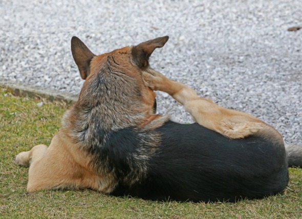
Your dog's activity will need to be restricted if the problem has been caused by a clotting disorder. A clot in a vein or artery can quickly become fatal when vigorous movement encourages the clot to travel to the heart. In cases of clotting, your dog will need to be treated specifically for dissolving the clot. In addition, if hyphema has significantly damaged your dog’s vision, it should not be allowed to go outside without supervision. Regular monitoring of the fluid pressure within the eye is also very important - daily checks for severe diseases, and in less severe cases, every two to three days until the condition has cleared up. To prevent your dog from inflicting further injury or irritation to the eye by scratching at it, you may want to ask your veterinarian for an Elizabethan collar - a wide collar that fits around the neck, preventing the dog from being able to reach its face with its paws.
Unless the ocular structures have suffered irreversible damage, the prognosis is usually good in case of traumas. In case of retinal detachments, secondary glaucoma will eventually develop, and surgical intervention may be necessary for relief of pain.

 Lung Cancer (Adenocarcinoma) in Dogs
Adenocarcinoma of the Lung in Dogs
Adenoca
Lung Cancer (Adenocarcinoma) in Dogs
Adenocarcinoma of the Lung in Dogs
Adenoca
 Hernia (Diaphragmatic) in Dogs
Diaphragmatic Hernia in Dogs
Diaphragmatic hernia
Hernia (Diaphragmatic) in Dogs
Diaphragmatic Hernia in Dogs
Diaphragmatic hernia
 Laryngeal Disease in Dogs
Disease of the Voice Box or Larynx in Dogs
The vo
Laryngeal Disease in Dogs
Disease of the Voice Box or Larynx in Dogs
The vo
 Liver Tumors in Older Dogs
Hepatic Nodular Hyperplasia in Dogs
Hepatic nodul
Liver Tumors in Older Dogs
Hepatic Nodular Hyperplasia in Dogs
Hepatic nodul
 'Mad Itch' Pseudorabies Virus Infection in Dogs
Suid Herpesvirus in Dogs
The pseudorabies virus i
'Mad Itch' Pseudorabies Virus Infection in Dogs
Suid Herpesvirus in Dogs
The pseudorabies virus i
Copyright © 2005-2016 Pet Information All Rights Reserved
Contact us: www162date@outlook.com