Normally, a heart contraction is caused by an electrical impulse originating from the sinoatrial node, stimulating the atria, traveling to the atrioventricular node and finally to the ventricles. This electrical conduction system is responsible for controlling the heart rate, and generating electrical impulses (waves), which propagate throughout the musculature of the heart, stimulating the heart's muscles to contract and push blood through the interior arteries and out into the body.
First-degree atrioventricular block is a condition in which the electrical conduction from the atria to the ventricles is delayed, or prolonged. On an electrocardiogram (EKG) this shows as a prolonged PR interval -- the time between the main electrical impulse, called the P wave, and the QRS complex, which is recognized as the heart beat.
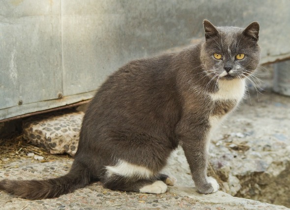
First-degree AV block may be found in young, healthy cats due to a high vagal tone (impulses from the vagus nerve that produce an inhibition in the heart beat), or it may be found concurrent with a degenerative conduction system disease.
Most cats with this condition will not display and symptoms. However, if induced by digoxin (heart medication) overdose, there may be a loss of appetite, vomiting, and diarrhea.
Although it may occur in otherwise healthy cats, those on certain prescription medications, such as digoxin, bethanechol, physostigmine, and pilocarpine, may be predisposed to first-degree AV block. Some other common causes for the condition include:
Low calcium, and certain drugs, may also predispose animals to first-degree AV block. First-degree AV block can also be caused by noncardiac diseases.
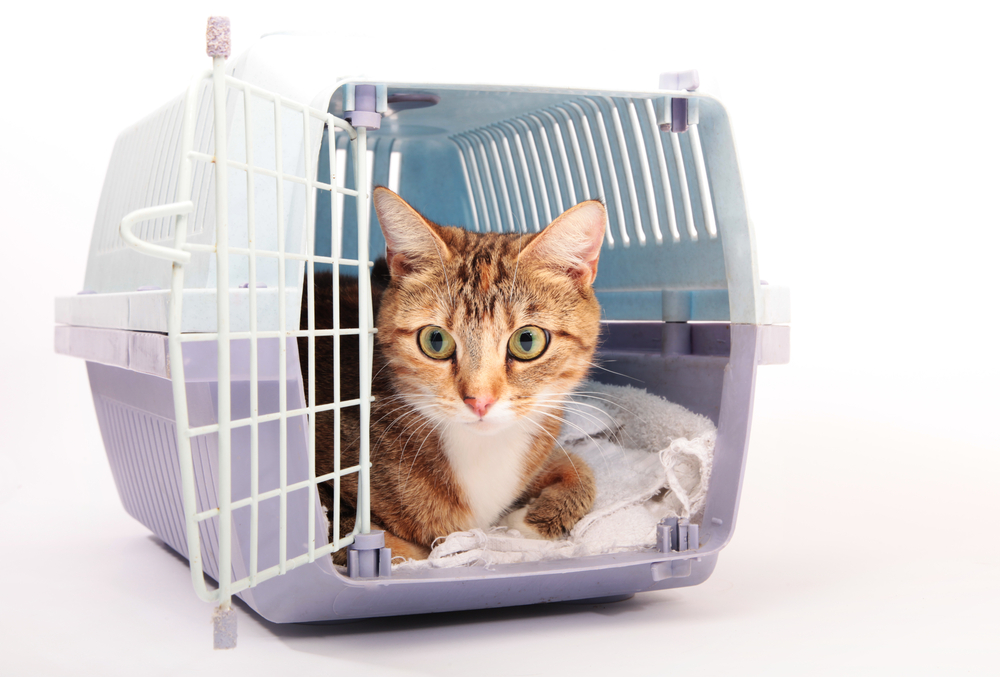
In addition to the full physical exam, your veterinarian will take a complete background history of your cat's health, when the symptoms began and any other symptoms that may point your doctor to the underlying cause. Standard tests include a chemical blood profile and a complete blood count to check for imbalances or infections.
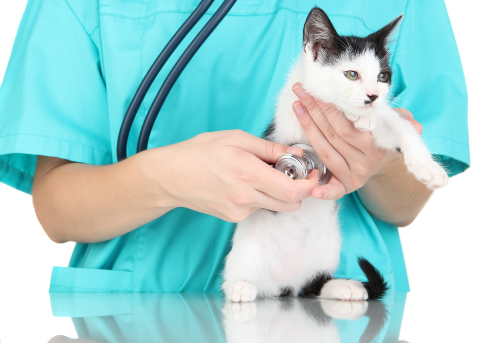
An echocardiogram (EKG) will be conducted to rule out certain types of heart diseases, and X-ray or ultrasound imaging can be used for internal imaging of the heart, confirming the presence of masses, or ruling them out. Gastrointestinal disorders, high pressure in the eye and upper airway disease are some of the diseases that can lead this disorder, all of which are unrelated (directly) to the heart.
An EKG recording can be used to examine the electrical currents in the heart muscles, and may reveal the exact abnormalities that are taking place in the cardiac electrical conduction (which underlies the heart’s ability to contract/beat).
Depending on the underlying disease causing the atrioventricular block, treatment will vary.
Your veterinarian will prescribe diet guidelines as necessary to treat the underlying cause of the disease. You will need to follow-up with your veterinarian for regular appointments so that any changes can be addressed immediately. EKGs will be taken at each visit to follow the progress of the heart's ability to conduct properly.

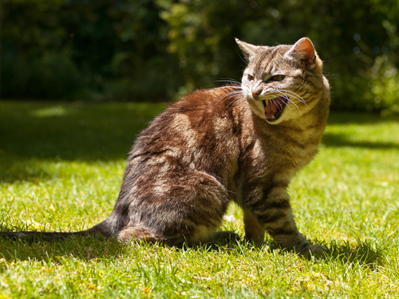 Aggression in Cats (Overview)
Cats are small, and often become the target of ot
Aggression in Cats (Overview)
Cats are small, and often become the target of ot
 Taurine Deficiency in Cats
Deficiency of Amino Acid Taurine in Cats
Amino ac
Taurine Deficiency in Cats
Deficiency of Amino Acid Taurine in Cats
Amino ac
 Kidney Enlargement in Cats
Renomegaly in Cats
Renomegaly is a condition in w
Kidney Enlargement in Cats
Renomegaly in Cats
Renomegaly is a condition in w
 Inflammatory Bowel Disease (IBD) in Cats
Inflammatory Bowel Disease (IBD) is a group of ga
Inflammatory Bowel Disease (IBD) in Cats
Inflammatory Bowel Disease (IBD) is a group of ga
 Chocolate Poisoning in Cats
Chocolate Toxicity in Cats
Although they’re not no
Chocolate Poisoning in Cats
Chocolate Toxicity in Cats
Although they’re not no
Copyright © 2005-2016 Pet Information All Rights Reserved
Contact us: www162date@outlook.com