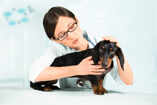
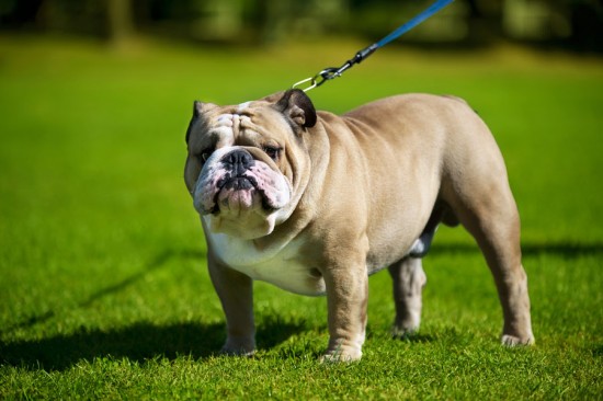
Hip dysplasia is a genetic disease which wears down the connective tissue, ligaments, and surfaces in the hip joint. In time, it causes arthritis and discomfort. Typically, it is diagnosed through a combination of tests. A complete physical exam is necessary, as well as an x-ray. The vet will also look for any signs of pain or arthritis. This can be seen through a simple examination, and will be revealed through an x-ray. The Orthopedic Foundation for Animals or OFA has developed a method which has set a standard which we still use today. The foundation was started in 1966. They are the largest all-breed registry in the world. Their method involves taking radiographs with specific guidelines. Usually, the minimum age of a dog before he or she is allowed to get an x-ray would be two years, because diagnosing hip dysplasia will be easier and more effective this way. Furthermore, those who are pregnant, nursing or in heat are not allowed getting an x-ray because subluxation happens to them during these times.
The OFA anesthetizes the dogs in order to relax the muscles ?important to get the best possible x-ray. Doctors and experts analyze the x-ray very carefully, looking through the congruity of the hip joints, the acetabulum, subluxation, the overall architecture (shape, size, etc.) of the head and neck of the femora. It takes three radiologists to see if the dog has dysplasia. They also rate if the dysplasia is mild, moderate or severe.
They are typically graded as either mild, moderate or dysplastic. Depending of the breed of the dog, the reliability of this diagnosis is around seventy to a hundred percent.
The University of Pennsylvania hip Improvement Program also has a unique method to diagnose hip dysplasia. They take radiographs at unique angles and views so they can see the amount of joint laxity, and can identify the disease more effectively. The program itself was established in 1983, and was first launched during 1993 as a fully usable system. Certification is required for all veterinarians, which can only be obtained through a special training course. This ensures the reliability and quality of the diagnosis. The system is applicable even to dogs as young as sixteen weeks.
The PennHIP heavily sedates the dogs before taking a radiograph. The radiographers take two views, when the position is neutral. This maximizes the laxity of the joint. These views are called distraction and compression radiographic views. Then, the ranges and distances are calculated. They quantify the displacement of the femoral head. They base their calculations on the distraction index. They take another radiographic view, a third, which makes use of the OFA抯 original radiographic view. This gives more information as it gives additional view of the femur.
This method, aside from being able to diagnose hip dysplasia even at an early age, is able to predict osteoarthritis. This is very helpful for those who want to prevent any diseases from getting worse through maintenance.
 What To Do If You Find A Stray Kitten
What To Do If You
What To Do If You Find A Stray Kitten
What To Do If You
 Mobile Grooming Services in Florida
Grooming y
Mobile Grooming Services in Florida
Mobile Grooming Services in Florida
Grooming y
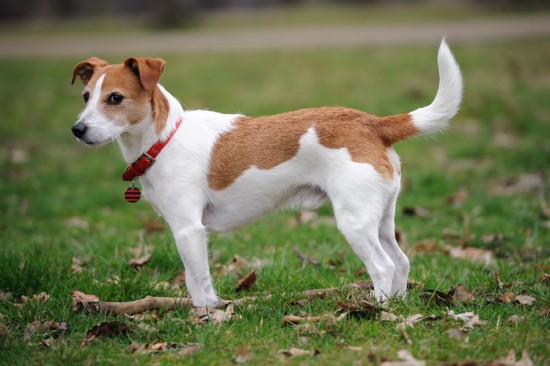 All About Terriers
All About Terrier
All About Terriers
All About Terrier
 Managing Hunting Behaviour In Dogs
Managing Hunting
Managing Hunting Behaviour In Dogs
Managing Hunting
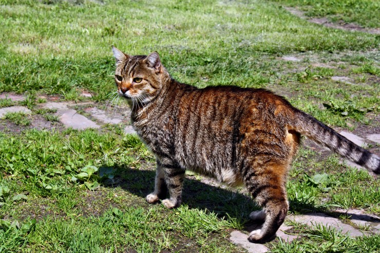 Tabby & Calico Shorthaired Cats, What’s The Difference?
Tabby & Calico Sh
Tabby & Calico Shorthaired Cats, What’s The Difference?
Tabby & Calico Sh