The spinal column is made up of a number of small bones called
vertebrae that are lined up like building blocks. A hole in the
center of each vertebra forms a tunnel in which the spinal cord
lies. The spinal cord is extremely important as it carries the
messages from the brain to the rest of the body. The spinal cord
is extremely delicate, and being surrounded by the bony
vertebrae helps to protect it. Between each pair of vertebrae,
just underneath the spinal cord, is a little cushion, called an
intervertebral disk. Disks cushion the vertebrae from one
another and provide flexibility to the spine during movement.
As a part of the normal aging process, these disks deteriorate,
resulting in so-called disk disease. Normally, each disk
consists of an outer fibrous ring and an inner gelatinous center
(a good analogy would be a jelly doughnut). With age this ring
becomes fragmented an the inner “jelly” center hardens to a
consistency of hard cheese. The fragmented outer ring may no
longer be able to hold this hard center in place, and movement
of the vertebrae on either side may suddenly squeeze the disk
out of its normal position. Unfortunately, this material usually
moves upward and comes to rest against the spinal cord, bruising
it in the process. This “slipping” of the disk often occurs
explosively, causing significant damage to the spinal cord and
pain to the animal. In this abnormal position the disk presses
against the spinal cord, causing further damage.
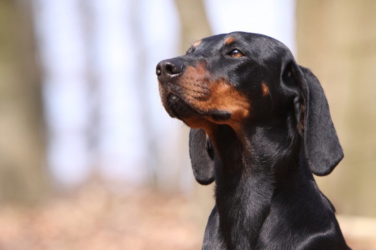
This type of disk disease may occur in dogs and cats of any age
or breed but occurs most commonly in the “short-legged” breeds
(e.g., dachshund, French bulldog, Welsh corgi, Pekingese) and
some other small breeds such as the poodle and cocker spaniel.
It may also occur in larger breeds of dog, including Doberman
pinschers. The parts of the spine most commonly affected by
“slipped” disks are the neck and the middle to lower back. When
a disk “slips” out of place and pushes against the spinal cord,
it usually causes the animal significant back pain and
frequently the damage to the spinal cord interferes with the
normal functions of the front and/or rear legs (depending on the
location of the disk rupture). In addition to being in pain, the
affected dog or cat may be lame, uncoordinated, and/or paralyzed.
These symptoms (pain, incoordination, and possibly paralysis)
indicate that the dog or cat has a problem affecting the spinal
cord but not the exact location or cause of the problem. Disk
disease, a tumor of the spine, or an infection of the spine may
all produce similar symptoms. Tests are needed to determine the
exact location and cause of the problem and to decide on the
appropriate therapy. In order to accomplish this, the patient
must be anesthetized for x-rays and collection of fluid from
around the spinal cord. “Myelography” is an x-ray study in which
a special dye is injected into the fluid surrounding the spinal
cord. This then allows any disk material pushing against the
cord to be identified on the x-rays. Analysis of the fluid
around the spinal cord helps to rule out other causes of the
problems such as infection.
In most cases disk disease is a problem requiring surgery to
remove the disk material compressing the spinal cord.
Occasionally, animals with disk disease are not treated by means
of surgery. In these animals, strict cage confinement and
immobilization are used. Usually this approach is used for a
first bout of back pain in animals that do not have problems
walking. Although strict cage confinement does not correct the
spinal cord compression, it may temporarily reduce some of the
pain and swelling around the spinal cord and permit the ruptured
disk to “heal.” As time goes on, it is not uncommon for animals
treated without surgery to suffer repeated bouts of pain,
lameness, and paralysis as additional disk material slips and
compresses the spinal cord. With each bout of disk disease the
spinal cord suffers additional permanent damage. Surgical
removal of disk material from the spinal canal is the only
treatment that provides rapid and maximal recovery of spinal
cord function.
Cortisone administration to animals with disk disease is of
therapeutic value only during the first 8 hours after the
initial spinal cord injury. Current scientific evidence does not
support the use of cortisone beyond this time. Furthermore, the
adverse effects of cortisone (e.g., stomach ulcers) must always
be kept in mind.
The surgery used most frequently to remove disk material from
around the spinal cord is called a laminectomy. For animals
undergoing a laminectomy, the speed of recovery and the extent
to which normal function of the legs is regained depend on many
factors, including the degree of the damage to the spinal cord
and the length of time that the spinal cord has been compressed
by the disk material. Animals exhibiting severe neurologic signs
(e.g., depressed feeling in their toes), a rapid onset of
symptoms (hours), and a long period of time before surgery
generally have a prolonged recovery period and may have varying
degrees of permanent damage.
The above is general veterinary information. Do not begin
any course of treatment without consulting your regular
veterinarian. All animals should be examined at least once every
12 months.
 Ferret Behaviour Decoded
 An Exercise In Exercise Patient: Why The Dog Is Banned From Future Workouts
 Choose ideal chicken houses and give your chicken the home it needs
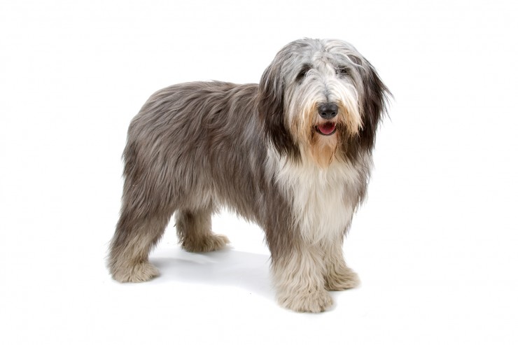 The Difference Between A Bearded Collie And A Polish Lowland Sheepdog
 Five Often Overlooked Financial Costs Of Cat Ownership
 The Gorgeous Austrian Black And Tan Hound
 Ten Great Ways For Pet Lovers To Help Animals This Christmas
Ten Great Ways Fo
Ten Great Ways For Pet Lovers To Help Animals This Christmas
Ten Great Ways Fo
 How To Make Your Dog The Star Of The Show
How To Make Your
How To Make Your Dog The Star Of The Show
How To Make Your
 How to Select Best Quality Foods for Dogs?
Dog
How to Select Best Quality Foods for Dogs?
How to Select Best Quality Foods for Dogs?
Dog
 Dealing With A Road Traffic Accident Involving A Dog
Dealing With A Ro
Dealing With A Road Traffic Accident Involving A Dog
Dealing With A Ro
 7 Of The Best Dog And Cat Treatment Hints !
7 Of The Best Dog
7 Of The Best Dog And Cat Treatment Hints !
7 Of The Best Dog
Copyright © 2005-2016 Pet Information All Rights Reserved
Contact us: www162date@outlook.com