Dystocia can be defined as inability to expel neonates through
the birth canal from the uterus. Dystocia is not uncommon in the
bitch and can have several causes. The diagnosis of dystocia
should be made and treatment instituted in an expedient fashion.
An incorrect diagnosis of dystocia may result in an unnecessary
caesarian section, but failure to recognize or prioritize
dystocia usually results in loss of puppies and perhaps even the
dam.
Dystocia can occur as a consequence of problems with the dam’s
uterus or birth canal or with the fetus. The diagnosis of
dystocia should be based on the presence of any of the following
criteria:
1. Failure of the dam to initiate labor at term. Bitches can be
considered over term at more than 70 to 72 days from the first
breeding, more than 58 to 60 days of diestrus, or more than 66
days from the luteinizing hormone (LH) surge or initial rise in
progesterone during estrus.
2. Failure of the dam to enter stage] labor beyond 24 to 36
hours after a detectable drop in rectal temperature to less than
99 to 1000F or to proceed from stage 1 to stage 2 labor within
24 hours.
3. Failure of the dam to complete delivery of all fetuses in a
timely fashion. Delivery should occur within 30 minutes to 1
hour of active labor (visible abdominal efforts) or 4 to 6 hours
of intermittent labor.
4. Fetal distress (unborn puppies with slow heart rates,
stillborns).
5. Maternal distress (excessive pain or systemic illness),
green or copious vaginal bleeding.
6. Irreversible history of dystocia (pelvic canal
abnormalities, mismatch between fetal and maternal size) or
radio-graphic evidence of fetal malposition.
Your veterinarian’s diagnosis of dystocia is based on taking an
accurate history, including reproductive history, ovulation
timing, and breeding dates, and performing a careful physical
examination including a digital pelvic examination for the
presence of vaginal abnormalities and the presence of a fetus in
the birth canal. A handheld Doppler device, abdominal
ultrasonography, and x-rays can be helpful in assessing fetal
viability, litter size, and fetal position. A blood test to
measure calcium and glucose levels may be helpful in identifying
metabolic disorders contributing to dystocia. Uterine
abnormalities contributing to the development of dystocia
include uterine inertia, abnormalities associated with fetal
fluids, and herniation or torsion of a uterine horn. Uterine
inertia, failure of the uterine muscle to contract in an
effective manner, can be primary or secondary. Primary uterine
inertia is multifactorial, with genetic, mechanical, hormonal,
and physical components.
Bitches exhibiting primary inertia fail to proceed into an
effective labor pattern, and cesarian section is indicated.
Bitches exhibiting secondary inertia fail to complete expulsion
of all fetuses because of exhaustion of the uterine muscle.
Medical management can be attempted, with adequate fetal
monitoring, but cesarian section may be necessary. Intravenous
glucose containing solutions and oxytocin (“pit”) and calcium
injections can be administered in appropriate doses.
Generally, minute doses of oxytocin are adequate (0.25 to 4.0
units per dog). Spastic, uncoordinated contractions of the
uterus occur if oxytocin is administered too rapidly or at too
high a dose. Uterine contractions interfere with fetal oxygen
supply by compressing placentas. Oxytocin should be administered
only with veterinary guidance. Abnormalities of fetal or
placental fluids include hydrops, an excessive accumulation of
allantoic fluid associated with each fetus, causing the fetal
unit to be markedly oversized. Rarely, underproduction of fetal
fluids occurs, resulting in dystocia caused by lack of
lubricating fluids.
Disorders of the birth canal contributing to dystocia include
pelvic abnormalities such as narrowing resulting from a healed
fracture or congenital disorders and vaginovulvar abnormalities
such as strictures. Successful natural breedings can occur
despite the presence of septate (vertical) bands in the vaginal
vault. Unfortunately, subsequent vaginal delivery of fetuses is
usually impaired. Strictures should be detected by the
veterinarian at the time of the soundness examination, before
breeding. Anular (circular) strictures are often detected at the
time of breeding, as they often interfere with the ability to
attain a natural tie. These should be repaired before breeding.
Bitches with unusually small vulvar openings may require a
partial episiotomy to deliver puppies vaginally.
Fetal causes of dystocia include fetal oversize; fetal
anomalies; and abnormal fetal position, presentation, or
posture. Fetal oversize can occur with prolonged gestation in
abnormally small litters (especially if there is a single pup)
and is the most common fetal cause of dystocia. Fetal anomalies
such as anasarca and hydrocephalus (abnormalities of body fluid
distribution) can cause a mismatch between the size of the birth
canal and that of the fetus.
Because both anterior (head-first) and posterior (breech)
presentations are normal in the bitch, only a transverse
(sideways) presentation is associated with dystocia and is rare.
Puppies are normally positioned with the fetal backbone adjacent
to the top surface of the uterus.
Malpositioning can cause mild dystocia. Abnormalities of
posture, normal being fully extended, are the second most
frequent fetal cause of dystocia. Malpositioning of the head,
forelimbs, or hindlimbs of the canine fetus is not readily
corrected with the use of forceps, traction, or digital
manipulation because of the limitations of the size of the birth
canal of the bitch.
The above is general veterinary information. Do not begin
any course of treatment without consulting your regular
veterinarian. All animals should be examined at least once every
12 months.

 Saint Bernard Dog Hereditary Health And Health Testing
Saint Bernard Dog
Saint Bernard Dog Hereditary Health And Health Testing
Saint Bernard Dog
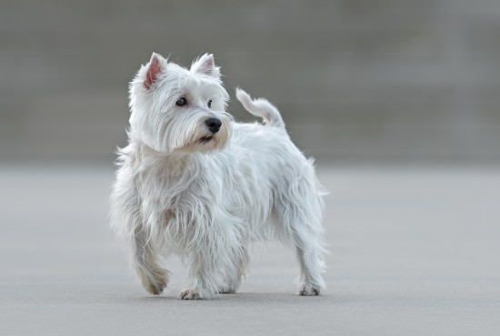 West Highland Terrier Skin Problems And General Health
West Highland Ter
West Highland Terrier Skin Problems And General Health
West Highland Ter
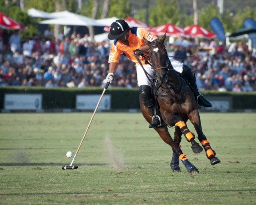 Introduction To Polo
Introduction To P
Introduction To Polo
Introduction To P
 Aspirin Poisoning In Dogs
Aspirin Poisoning
Aspirin Poisoning In Dogs
Aspirin Poisoning
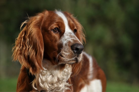 How To Keep A Welsh Springer Spaniels Coat Looking Good
How To Keep A Wel
How To Keep A Welsh Springer Spaniels Coat Looking Good
How To Keep A Wel
Copyright © 2005-2016 Pet Information All Rights Reserved
Contact us: www162date@outlook.com