Cardiomyopathy is the name applied to an abnormality of heart
muscle function. The heart’s pumping ability is diminished,
resulting in such signs as inability to exercise; fatigue;
fainting; fluid collection in the lungs, abdomen, and limbs; or
emboli (clots that arise in the heart and travel to the kidney,
brain, or legs). Although some patients with cardiomyopathy do
not develop clinical signs, others experience rapid progression
of their disease or sudden death. The causes of cardiomyopathy
include genetic predisposition, infections, toxic causes (drugs
and chemical compounds), specific dietary insufficiencies, and
unknown causes. Whereas some cases are entirely reversible,
others are not and are treated with various levels of success.
Three major forms of cardiomyopathies occur in the canine and
feline species. In dilated cardiomyopathy, the heart muscle is
weak and flaccid (floppy). This condition is associated with a
reduction in heart muscle function during contraction (systole)
and a decrease in forward flow of blood. Subsequent upper heart
chamber (left atrial) enlargement is associated with backup of
blood and then fluid into the lungs (pulmonary edema).
Hypertrophic cardiomyopathy is a thickening of the lower heart
muscle chambers (ventricles). The results are inappropriate
heart function, obstruction of blood flow from the heart into
the circulation, and enlargement of the upper heart chambers
(atria). This abnormality is called diastolic dysfunction, a
condition in which the heart fails to relax fully, fill, and
then empty. The resulting backup of pressures into the lung is
responsible for the clinical signs of respiratory distress,
coughing, and systemic emboli (blood clots).
Unclassified or restrictive cardiomyopathies are unidentified
disease conditions in which heart problems are associated with
severely enlarged upper chambers and diminished pumping ability.
The clinical signs resemble those of hypertrophic
cardiomyopathy. Although not thickened, the ventricular muscle
is dysfunctional and the heart is unable to fill and then pump
adequately.
Cardiomyopathies are seen in both dogs and cats. The form in
dogs is usually dilated, whereas hypertrophic and unclassified
forms are identified most often in cats. The diagnosis of
cardiomyopathy is based on a history of weakness, coughing,
panting, fainting, or fluid collection around the lungs and in
the abdominal cavity. Weight loss occurs, and seizures
associated with fainting may occur. Emboli (clots) can result in
blood vessel blockage, sudden lameness, and cold painful limbs.
Clinical signs usually develop suddenly, often without apparent
prior illness. In addition to these signs, the diagnosis depends
on abnormalities found at the physical examination.
Irregularities occur in the heart’s rhythm and rate, and
abnormal heart sounds (murmurs) are heard with the stethoscope.
Radiographs (x-rays) of the chest show heart enlargement.
Evaluation of the blood may identify complicating organ
problems. The electrocardiogram can diagnose and irregular heart
rhythm and substantiate heart enlargement. Ultrasound
examination of the heart confirms the suspicion of
cardiomyopathy. Dilation of the heart cavity, poor contractility
of the heart muscle, and left atrial enlargement occur with
dilated cardiomyopathy. Thickening of the heart muscle,
obstruction of the flow of blood into the circulation, and left
atrial enlargement identify hypertrophic cardiomyopathy. Normal
muscle thickness with disturbed function and enlarged left atria
indicates restrictive cardiomyopathy.
Treatment varies with the type of cardiomyopathy. Dilated
cardiomyopathies, indicative of a loss of contractile heart
strength, require medications to improve strength (digitalis),
to remove excess fluid accumulation (diuretics), and to
counteract abnormal hormone levels that contribute to heart
failure (angiotensin-converting enzyme inhibitors). A low-salt
diet is important to reduce sodium levels and subsequent water
retention. Nutrients such as taurine and carnitine may be
required to counteract specific deficiencies. Manual removal of
excess fluid accumulation is sometimes necessary. Treatment of
hypertrophic and unclassified cardiomyopathies requires drugs to
allow the ventricular muscle to relax. This improves heart
filling and blood flow to the body. Beta-adrenergic blocking
agents or calcium channel blocking agents are often used for
this purpose. Removal of excess fluid from the body (diuretics)
and sometimes manual removal of fluid from the chest space are
necessary to improve comfort.
Low-salt diets to counteract salt and water retention are
indicated but may be difficult to achieve with a finicky and ill
cat. Aspirin is used to reduce the likelihood of blood clot
formation within the heart. Antiarrhythmic agents to control
irregularities of the heart’s rate and rhythm are called upon at
times, as are nutritional supplements (taurine and/or carnitine)
in known deficiencies. The prognosis for survival with
cardiomyopathies varies from poor to good. Once cardiomyopathy
has been recognized, much of the damage to the heart muscle has
already occurred. The result is congestive heart failure, the
signs and symptoms of which may be treated for a variable period
of time (often 3 to 12 months, which is equivalent to 3 to 5
years in a human).
Although the pet may enjoy a period of good health and comfort,
the long-term prognosis continues to indicate that heart failure
will recur. As a result, the pet will become less responsive to
medical intervention. Surgery is not yet an option for any form
of cardiomyopathy.
The above is general veterinary information. Do not begin
any course of treatment without consulting your regular
veterinarian. All animals should be examined at least once every
12 months.


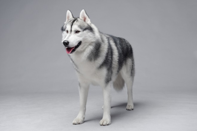
 Vulnerable Native Uk Dog Breeds - The Hound Group
Vulnerable Native
Vulnerable Native Uk Dog Breeds - The Hound Group
Vulnerable Native
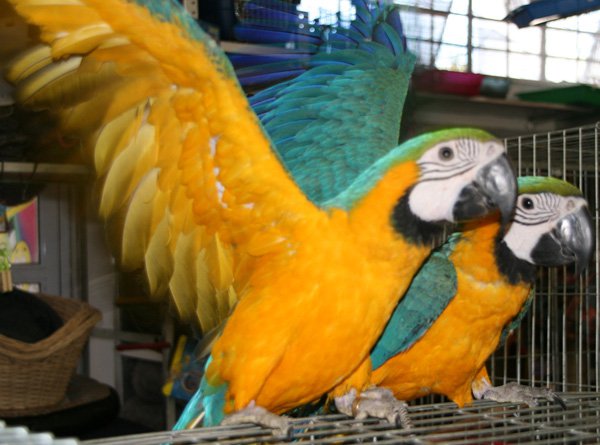
 April Is National Pet Month – Celebrate With Us!
April is National Pet Month! You may be asking yourself, Wha
April Is National Pet Month – Celebrate With Us!
April is National Pet Month! You may be asking yourself, Wha
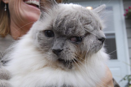 What Is A Janus Cat?
What Is A Janus C
What Is A Janus Cat?
What Is A Janus C
 Tracheal Collapse In Dogs
Tracheal Collapse
Tracheal Collapse In Dogs
Tracheal Collapse
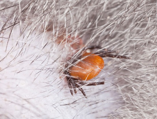 How To Identify And Remove Ticks From Your Pet
How To Identify A
How To Identify And Remove Ticks From Your Pet
How To Identify A
Copyright © 2005-2016 Pet Information All Rights Reserved
Contact us: www162date@outlook.com