Hypertrophic osteodystrophy or HOD is a condition that affects young large breed dogs. HOD is a bone disease that affects the rapid growing bones of giant dog and may occur between ages of 2 and 7 months. The breeds that are at high risk for HOD are Boxers, Chesapeake Shepherd Dog, Golden Retriever, Great Dane, Irish Setter, Labrador Retriever and Weimaraner, although there can be exceptions to this rule. Commonly, large male dog breeds are more affected than females. Inherited or genetic link seen to be affects in occurrence of this disease.
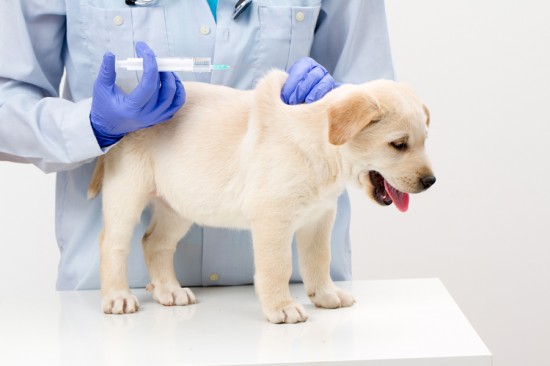
There is currently unknown or no agreement on the cause of Hypertrophic osteodystrophy. Possible causes maybe considered are; bacterial infections, canine distemper virus infection, vaccination with distemper virus or any other viral infections. Also Vitamin C deficiency is also speculated, hence the decreased uptake of Vitamin C and/or increased uptake of other vitamins and minerals other than vitamin C. The excessive calcium supplementation is also included as one possibility. There may be a link to recent vaccination with a modified live vaccine, but no specific vaccine has been implicated. on Weinmaraner dog breeds, it is recommended for them to receive killed virus vaccines instead of modified live or separate vaccines for canine distemper, parvovirus, and adenovirus to prevent the possibility of vaccine-induced HOD.
The signs and symptoms of HOD appear to be mild to moderate painful swelling of the growth plates in the leg bones of dogs. It most commonly affects the ends of the radius, ulna (the long bones from the elbow to the wrist) and tibia (the long bone from the knee to the hock). Lameness may vary from mild to sever, reluctance to stand if multiple limbs are affected. Fever, anorexia, loss of appetite and depressions are noticed. Swelling and heat are commonly present over the affected bones. Some clinical signs also includes diarrhea, discharge from the eyes, tonsillitis, thickening of the foot pads, pneumonia, and abnormal development of the enamel of the teeth. Dogs suffering really proper petsafe and care.
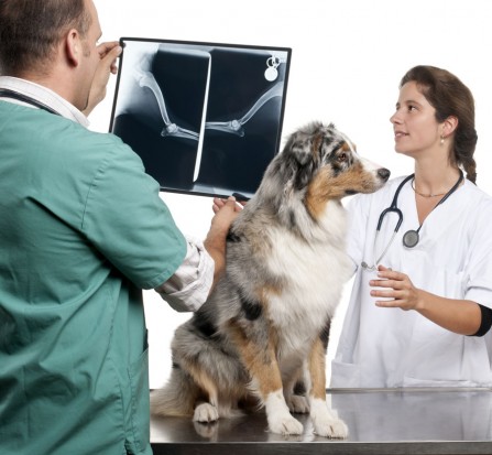
X-ray signs of HOD are more clearly noticed. A line of lucency where the bone has been destroyed is usually found to be parallel to the growth plates of the affected bones. X-rays show a dark line at the metaphysis, which can progress to new bone growth on the outside of that area. This represents microfractures in the metaphysis and bone proliferation to bridge the defect in the periosteum. Some signs seen on microscope are also clear. The growth plate is normal, but blood vessels adjacent to the growth plate are frequently dilated. Bleeding in the bone adjacent to the growth plate and extensive death of the bone adjacent to the growth plate. Adjacent to the line of lucency is a zone of increased density of bone that corresponds to collapsed of layers of dead bone. The outer layer of the bone (periosteum) is thickened with new bone formation.
The treatment for HOD is generally supportive since this is a very painful condition and these disease is usually self-limiting which can last a few weeks. Treatment includes intravenous fluid therapy, anti-inflammatories and painkillers such as buffered aspirin or carprofen (Rimadyl) are given and needed enough rest on their comfortable pet beds. In addition, the animals are usually given a broad-spectrum antibiotic since bacterial infection is suspected. Since the dog might be too irritable and uncomfortable, strict rest on a comfortable warm bed is recommended. Feeding a nutritious, highly palatable food will help to encourage some dogs to eat. In severe cases steroids may need to be given to control the pain, but because of the possibility of this being a bacterial disease their use may be contraindicated due to their immunosuppressive qualities. Supplementation of Vitamin C is contraindicated due to an increase in calcium levels in the blood, possibly worsening the disease. Permanent skeletal deformity can occur, recurrence can be a problem until the dog reaches maturity and dogs usually do not die of the disease rather are euthanatized if recovery is poor or if clinical signs are severe.
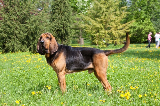 10 Very Talkative Dog Breeds
10 Very Talkative
10 Very Talkative Dog Breeds
10 Very Talkative
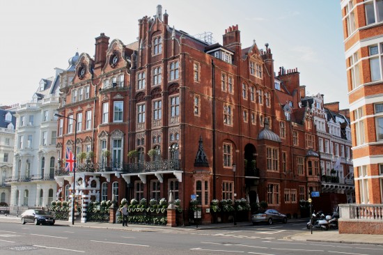 5 Luxurious Dog Friendly Establishments Dotted Around The Country
5 Luxurious Dog F
5 Luxurious Dog Friendly Establishments Dotted Around The Country
5 Luxurious Dog F
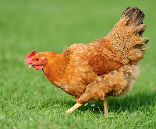 Healthy Skin And Feathers In Chickens
Healthy Skin And
Healthy Skin And Feathers In Chickens
Healthy Skin And
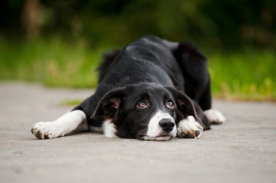 Recognising When A Dog May Be Suffering
Recognising When
Recognising When A Dog May Be Suffering
Recognising When
 Regular Pet Grooming Is Crucial
Pet owners hav
Regular Pet Grooming Is Crucial
Regular Pet Grooming Is Crucial
Pet owners hav
Copyright © 2005-2016 Pet Information All Rights Reserved
Contact us: www162date@outlook.com