Stem cell implantation: what exactly is it? Some pet owners may initially be unsure about stem cell procedures and what they entail, but at our animal hospital in McKinney, TX, we've found that pet owners usually opt for the procedure once they understand how safe and effective it is.
Stem cell implantation is currently recommended in animals for osteoarthritic conditions (most notably hip dysplasia) and disorders involving tendons and ligaments (most commonly partial tears of the cranial cruciate ligaments).
Studies are underway to assess feasibility of treating a host of other conditions including renal and liver diseases, autoimmune disorders, and chronic allergic dermatoses. In fact, our animal hospital in McKinney, TX is seeking cats with early renal failure to treat with stem cells, which research has shown can halt the progression of renal cellular failure.
Stem cell implantation must be considered only for animals without signs of cancer or other disorders such as active infection. A physical examination and a blood profile are done to search out the presence of coincidental disease that must be addressed before implantation. We also take chest radiographs to search for any evidence of metastatic cancer in the lung tissue.
Having completed our examinations and found the patient a suitable candidate for stem cell treatment, the next step is resecting fat tissue to submit for stem cell harvesting. There are several sites on the body to obtain fat tissue, including the falciform ligament, greater omentum (a lace-like tissue that encompasses the intestines), bone marrow, a region just behind the shoulder blades, and from the inguinal area between the upper rear legs and body wall.
The falciform ligament is the most favored source of fat; it usually provides abundant samples that yield the greatest numbers of cells. This is located on the midline just forward of where a spay incision is located. The procedure requires general anesthesia and involves an approximately 3-inch incision. Since this gives us access to the abdominal cavity, we're able to remove some greater omentum in those patients (usually very small dogs or cats) that have insufficient fat deposits in the falciform ligament.
Recovery from this procedure is rapid and patients are released later that day with medication for pain relief and antibiotics.
Very strict attention is paid in all steps to assure complete sterility of the fat. It's transferred to vials that are overnight-shipped to the VetStem labs in California. There, mesenchymal stem cells are derived and their sterility confirmed before shipment and delivery back to our hospital two days later. Though we can delay shipment to accommodate the pet owners' scheduling convenience, we usually schedule the patient to be back in two days for implantation.
Cells usually arrive at around 10:00 in the morning. An intravenous catheter is placed and the patient is prepped for sterile surgery. Every conceivable effort is made to ensure absolute sterility at every moment.
The procedure involves injections of stem cells within affected joints along with 5 CC of cells given intravenously. We have placed cells within hips, elbows, knees, and hocks (ankles) for our patients. For treatment of bilateral hip dysplasia, for example, the hip areas are clipped and then scrubbed with surgical cleanser and sprayed with iodine solution. Draping is done to isolate the area to be injected.
At our animal hospital in McKinney, TX, we place a sterile needle into the hip joint, and then inject the joint with cells. The patient is re-positioned to allow access to the other hip, and the procedure repeated. At this point cells are injected intravenously to conclude the implantation.
The joint can be sore for a few days after the implantation because of the needle passing through the joint capsule and because of the manipulations used to ensure joint access. We prescribe pain medication to control this discomfort, and sometimes for prolonged periods of time when joints are very painful due the original condition. Antibiotics are also prescribed as a further measure to assure asepsis of the joint environment.
We recommend exercise restriction for one month following the treatment, limited to leash walks without vigorous activity. Range of motion exercises are also begun. Sutures from the original surgery are removed in 10 days, which gives us a chance to evaluate progress and assess for joint function. Our patients are often surprisingly improved at that recheck, and pain medication is weaned off accordingly.
When compared to surgical procedures, stem cell therapy is a safe, relatively painless, and cost-effective method to relieve chronic pain from arthritis and ligament/tendon disorders. Our animal hospital in McKinney, TX has stopped offering hip replacement surgery, for example, because patients do so well (even months and years later) without major orthopedic procedures.
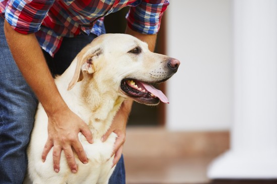 Do Owners Influence A Dogs Behaviour?
Do Owners Influen
Do Owners Influence A Dogs Behaviour?
Do Owners Influen
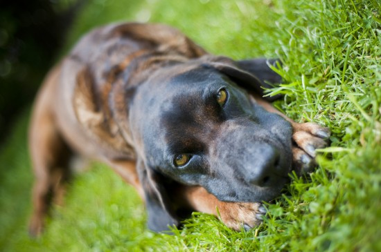 Health Issues More Commonly Seen In The Bavarian Mountain Hound
Health Issues Mor
Health Issues More Commonly Seen In The Bavarian Mountain Hound
Health Issues Mor
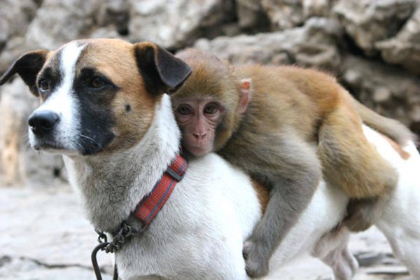 Keep your dog warm in winter!!
In the event th
Keep your dog warm in winter!!
Keep your dog warm in winter!!
In the event th
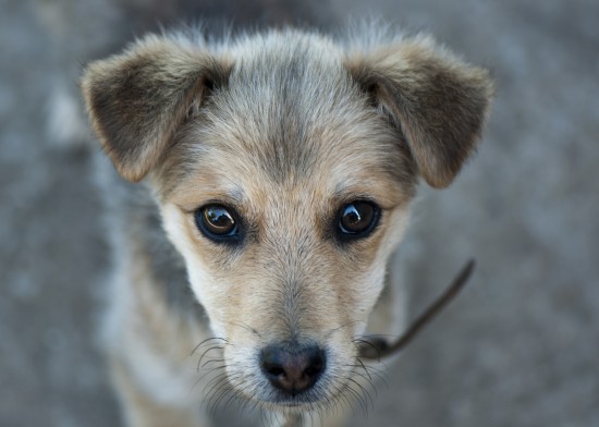 Gaining The Trust Of An Abandoned Or Mistreated Puppy
Gaining The Trust
Gaining The Trust Of An Abandoned Or Mistreated Puppy
Gaining The Trust
 The Origins Of Our Feline Friends - Cats
The Origins Of Ou
The Origins Of Our Feline Friends - Cats
The Origins Of Ou