When the veterinarian looks into your pet’s eyes, he or she isn’t just checking the animal’s vision. The eyes are the perfect window into your furry friend’s state of health, and can reveal medical problems in addition to compromised vision.
The ophthalmic examination generally accompanies a very thorough physical examination. If your pet is sick from a systemic disease, often times the eyes will give your veterinarian clues that something is wrong before any diagnostic tests are run.
The eyes are the perfect window into your furry friend’s state of health.
The beginning of the ophthalmic exam involves basic observation of your pet’s ability to navigate around the examination room.
The lights often are turned down to ensure that the patient can see in darker light as well. Vision is also roughly evaluated by doing something called the “menace” test: the veterinarian will make a menacing movement with his or her hand in front of the eye being evaluated without hitting any facial hair or whiskers. If the eye is healthy, then the animal should blink or move away from the movement. Another common test is to drop or toss a cotton ball in front of the eye to see if the pet responds by watching it fall.
In addition, the veterinarian most likely will run a test called a pupillary light reflex, or PLR, to evaluate the retina, cranial nerves, the muscles controlling the iris and part of the brain involved in visualization. This test requires the use of a very bright focal light, which will be shined into one eye at a time to make sure that the iris closes down and the pupil constricts normally. The eye that does not have the light shined into it is also evaluated to be sure that pupil constricts as well.
If the veterinarian feels it is necessary, he or she may also conduct a Schirmer tear test. With this diagnostic procedure, small paper test strip is placed in each lower eyelid and left in place for 60 seconds, and then removed. The veterinarian is then able to measure the amount of tear production in that eye. Veterinarians run these tests because tears are responsible for helping to keep the eye healthy, moist and free from infection. A deficiency in tear production often means the eye has keratoconjunctivitis sicca or dry eye. If left untreated, this condition can lead to blindness.
As your pet’s guardian, it is very important that you stay on alert for changes in your pet’s ability to see.
The veterinarian probably will assess the upper and lower eyelids and conjunctiva to be sure that there are no abnormalities such as infection, inflammation, a foreign body, or a growth or mass. Both the upper and the lower eyelids contain tiny openings called puncta that allow tears to drain from the eyes down into the nose. This passageway is referred to as the nasolacrimal drainage apparatus. If it becomes blocked for whatever reason, then tears spill off the eyelid and often stain the hair below the eyes.
To determine if the drainage system is open and allowing the tears to flow normally, a stain known as fluorescein dye is placed into each eye. The stain should turn apple-green and if the duct is working properly, the dye will be seen dripping out of the nostril on the same side as the eye treated with the dye. If the passage is blocked, then the veterinarian can flush the ducts to reopen them. This treatment is referred to as a nasolacrimal flush.
Placement of the fluorescein dye will also determine if the cornea or the clear covering of the eye is normal and healthy. If the dye adheres to the cornea, the eye has a corneal ulcer or defect in the epithelium. Corneal ulcers must be treated immediately or they can become infected and lead to permanent vision loss. The cornea will be evaluated for any other abnormalities, such as blood vessels, pigment or other defects that may indicate another problem that needs treatment.
The next part of the examination involves evaluation of the interior part of the eye or the part behind the cornea. Using an ophthalmoscope, the veterinarian can determine if there is an inflammation of the anterior uvea, a condition referred to as uveitis. The lens of the eye is also evaluated for abnormalities, such as cataracts, which can cause blindness.
An ophthalmoscope is useful, as well, for examining the back of the eye or the ocular fundus, a part of the eye that includes the retina and its blood vessels, as well as the optic nerve head. The veterinarian will check the retina for any changes that could lead to blindness such a progressive retinal atrophy, late-onset retinal degeneration that can occur in older dogs, and retinal detachment. The blood vessels of the retina are also evaluated. Animals suffering from systemic diseases such as hypertension from kidney disease or hyperthyroidism have changes in the retinal blood vessels that can be seen on examination.
Following evaluation of the retina and structures inside the eye, the veterinarian may need to determine the intraocular pressure or the pressure within the eye. A higher-than-normal pressure is indicative of glaucoma; a lower-than-normal pressure might indicate an inflammation of the eye or uveitis. It is important to check the pressure of the eyes if the animal is from a purebred line that is at increased risk for developing glaucoma or other genetic eye diseases. A few canine breeds that seem to be predisposed to developing glaucoma include the Akita, beagle, Bouvier des Flandres, chow chow, golden retriever, Great Dane, Norwegian elkhound, samoyed, and Siberian husky.
As your pet’s guardian, it is very important that you stay on alert for changes in your pet’s ability to see, as well as any signs that your cat’s or dog’s s eyes seem to be causing him or her discomfort. If your pet has been bumping into objects, has shown difficulty or hesitance in using the stairs, or an inability to get around in a new environment, notify your veterinarian immediately: the animal’s vision may be at stake. Signs of discomfort within the eyes include pawing or rubbing of the eyes, tearing or squinting, hiding from the light, the presence of discharge from the eyes, or redness. In addition, if you notice a bluish haze or change in color of the cornea, swelling of the eyeball or lids, or any other change in your pet’s eyes, you should notify your veterinarian right away.
If your veterinarian suspects that something is wrong with your pet’s eyes and he or she suggests that you seek a second opinion or advanced specialized care, then a referral to a veterinary ophthalmologist can be arranged. An ophthalmologist has the expertise and equipment to handle more complicated medical problems with your pet’s eyes.
 Why You Should Adopt A Rescue Dog For Your Family
Credit: Flickr: Ian Phillips
Why You Should Adopt A Rescue Dog For Your Family
Credit: Flickr: Ian Phillips
 How To End The Fear Of Fireworks For Your Dog
Dogs And FireworksHaving bee
How To End The Fear Of Fireworks For Your Dog
Dogs And FireworksHaving bee
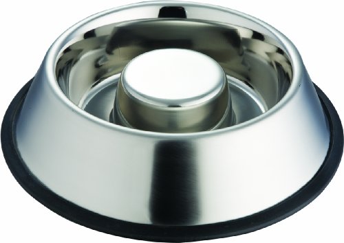 Indipet Slow Feed Pet Bowl
If you have a dog that loves
Indipet Slow Feed Pet Bowl
If you have a dog that loves
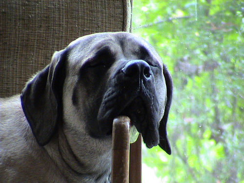 10 Dog Breeds For Laid Back Lifestyles
Make Room On The
10 Dog Breeds For Laid Back Lifestyles
10 Dog Breeds For Laid Back Lifestyles
Make Room On The
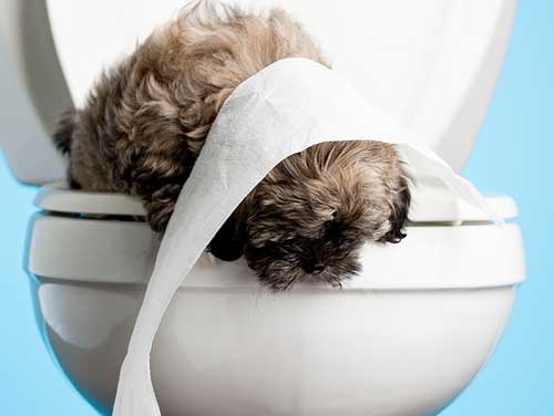 How to potty train your puppy
Dont break the house rulesPotty train your dog
How to potty train your puppy
Dont break the house rulesPotty train your dog
Copyright © 2005-2016 Pet Information All Rights Reserved
Contact us: www162date@outlook.com