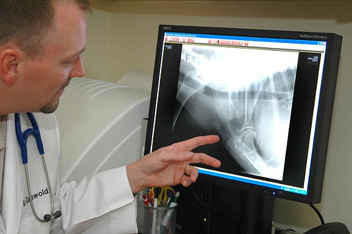
By T. J. Dunn, Jr., DVM
Bone cancer in dogs and cats can be a challenging disorder to overcome. Although rare in cats, bone cancer in dogs is most common in large breeds but can occur in any canine. Achieving and maintaining good health is a balancing act.
There is an invisible chemical ebb and flow, a true harmonic resonance that lives within a healthy animal. And when that vibrant harmony is upset, when the sweet song of life is bumped out of balance, ill effects sweep over the entire individual. Cancer is one notable form of disharmony within an individual.
The hallmark of cancer is uncontrolled cell growth, invasion of cells into nearby structures and sometimes a dispersal to distant organs, which is termed metastatic cancer. And since any cell in the dog’s body has the potential to develop into a cancerous cell, bone cancer dramatically illustrates what can happen when things go wrong.
When a cell turns cancerous by a disruption the cell’s physiology, structure or function, normal neighboring cells usually consume the rogue cell. On other occasions the defective cell simply self-destructs and is swept away. But when conditions are just right -- or wrong from the animal’s perspective -- a modified cell, called a mutant, survives the modification, retains its vigor and reproduces more cells just like itself.
Generation after generation of cells arising from that single mutated cell eventually changes the neighborhood and carves out it’s own territory, spreading its own bad seeds into more and more neighborhoods. Metastatic bone cancer cells break away, hitch hike the blood stream or lymph fluid and travel to entirely new neighborhoods within the dog’s body and begin the malignant process all over again.
Cancer is also termed neoplasia, which means new growth. A cancerous cell grows faster than normal and divides and multiplies at an abnormal rate; its progeny do likewise. From that one abnormal neoplastic cell more like itself invade and crowd out surrounding tissues. With bone cancer, there are four types of cell lines capable of evolving into a neoplastic condition:
 1. Osteosarcoma … causing nearly 80 percent of all bone cancers this most common form of bone cancer arises from cells that deposit bony minerals. Aggressive invasion and rapid growth make this form of cancer a dreaded threat. The X-ray image to the right shows what an osteosarcoma of the humeral head looks like (click on it to enlarge).
2. Chondrosarcomas … these tumors arise from the cartilage joint surfaces at the ends of bone and generally have a less aggressive tendency to invade and spread.
3. Fibrosarcomas … originate from fibrous connective tissue adjacent to bone, are locally invasive into the bone and have a low tendency to spread.
4. Synovial cell carcinomas ... originate from joint tissues and invade the associated bone. These tumors are less aggressive than osteosarcomas.
A definitive diagnosis of bone cancer can only be made via microscopic evaluation of a bone biopsy. Veterinary pathologists classify the degree of malignancy of the cells and likeliness of metastasis to other tissues. Like seeds on the wind, neoplastic cells can be carried by the blood and lymph from the original site of the cancer to distant tissues whereupon a new cancerous growth arises. Called metastatic cancer, whenever distant growths are present in a dog’s body the magnitude of the ill effects on the patient are remarkably increased -- and the chances of a cure drastically reduced.
Most commonly seen in long bones such as the femur, bone cancer has a predilection for larger breeds including the Greyhound, Saint Bernard and Mastiff. Chronic, low-grade lameness with gradually increasing swelling near a joint will alert the veterinarian to the potential for a tumor to be present. X-rays of the affected area often display characteristic changes in a bone that are totally unlike the defects usually associated with arthritis.
On occasion, an apparently normal dog will be presented with a spontaneous, severe lameness. Physical examination and radiographic evaluation, to everyone’s shock, reveals the cause of the break to be due to bone cancer. This break is termed a pathological fracture and there is an example of a pathological fracture in the table below.
Osteosarcoma continues to be one of the most challenging types of cancer to treat. Part of the therapeutic challenge arises from the fact that at the time of diagnosis there often has already been metastasis to other areas of the body.
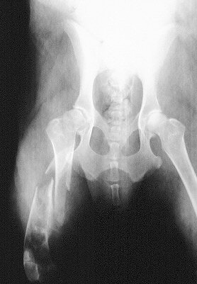
 This image is of a pathological fracture of the femur that was caused by cancer of the bone weakening the bone structure
This closer side view of the pathological fracture is of the patient on the left
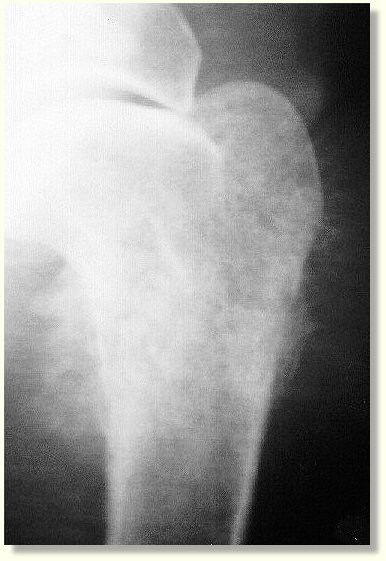
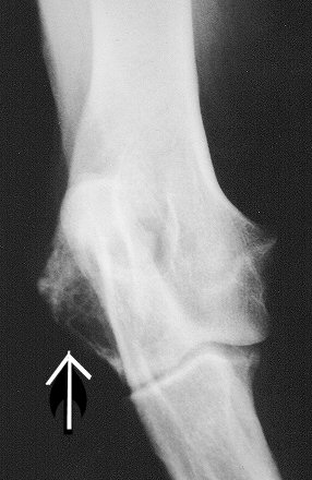
The arrow points to bone cancer invading the lateral side of the humerus near the elbow joint
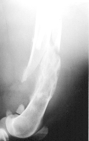
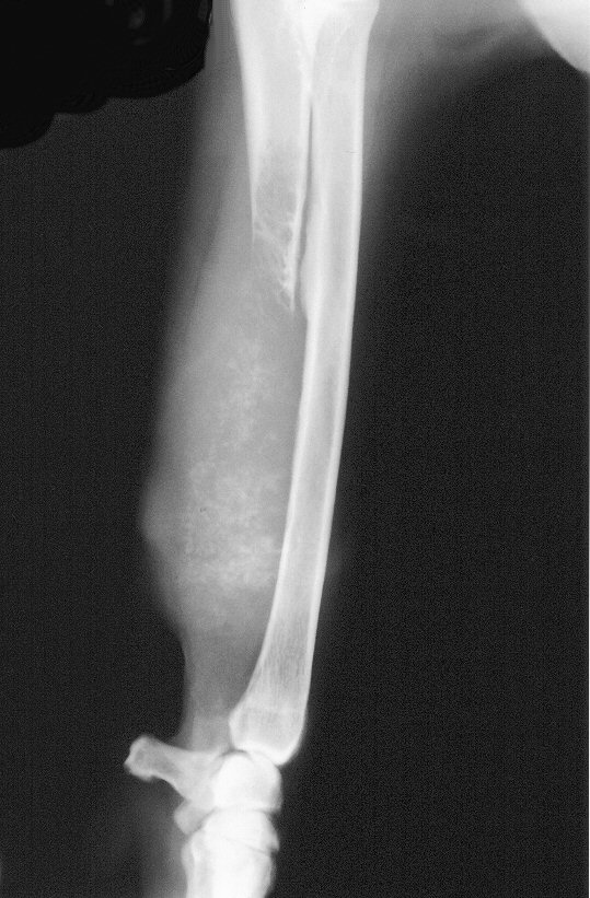
This image displays extreme dissolution of the distal two thirds of the ulna of a dog due to bone cancer
This image is of a pathological fracture of the femur that was caused by cancer of the bone weakening the bone structure
This closer side view of the pathological fracture is of the patient on the left
The arrow points to bone cancer invading the lateral side of the humerus near the elbow joint
This image displays extreme dissolution of the distal two thirds of the ulna of a dog due to bone cancer
"Unfortunately, at the first signs of lameness," says Dr. Kenneth M. Rassnick, Assistant Professor of Oncology at Cornell University College of Veterinary Medicine, "we expect that the tumor has already metastasized. However, as long as the metastatic cells are still microscopic and we cannot detect them on radiographs, then dogs will still benefit from treatment."
There is no single treatment protocol for all patients with bone cancer; Rassnick explains that individualized strategies are selected for each patient. "Currently, for dogs with osteosarcoma, I thoroughly screen them for obvious signs of metastasis. For most dogs, this includes radiographs of the lungs and physical examination and palpation of other bones. Amputation of the affected leg is the first line of treatment but unfortunately, amputation alone is only palliative for a cancer as aggressive as osteosarcoma. Over time, the metastatic cells will continue to grow in number and size. If it is determined by radiography, ultrasound or physical exam that there are no metastatic tumors present, amputation of the affected leg followed by chemotherapy has proved to be the most effective treatment for osteosarcoma. There are a number of chemotherapy regimens that we know to be effective at controlling the metastatic cells."
Thoughtful consultation with the veterinarian regarding chemotherapy is very important. Rassnick tells us "The exact chemotherapy protocol will depend on a number of factors including overall health status of the dog and function of organs such as the heart and kidneys. We have spent a considerable amount of time trying to determine the best time to begin chemotherapy. Since we know the cancer cells have already spread, it is tempting to begin therapy as soon as possible. Some have even advocated giving chemotherapy before the amputation surgery or even at the same time. Our studies have shown there is not a tremendous benefit to instituting therapy that soon, so I generally recommend having the amputation surgery done, letting my patients heal for 7-14 days and then begin chemotherapy when the stitches are ready to be removed."
Not all dogs will be candidates for amputation. Rassnick adds "Some dogs may have concurrent orthopedic or neurologic problems that may complicate ambulating with three legs, or occasionally it is the wish of the family not to pursue the surgery. We can offer palliative options for controlling bone pain including nonsteroidal medications and even radiation therapy. Localized radiation therapy to the diseased bone is often a very effective method at controlling the pain and some veterinary oncologists can now offer this as an option."
As drastic as amputation seems to be, it should not be immediately rejected as an attempt at treatment. As a practitioner for over thirty years I have been amazed at how some canine amputee patients respond and adapt. Each case should be evaluated on its own merits with attention paid to such factors as the presence of arthritis in the patient, degree of excess body weight, heart and other organ function, and the patient’s attitude and ability to adapt to new situations.
Until new research reveals more about the menace of cancer’s mysterious origins and until ways are found to turn off rapidly multiplying cancer cells, we will need to be on the alert for bone cancer in our dogs. A veterinarian should evaluate any lameness that persists longer than three days. All dog owners should be proactive in requesting that an x-ray evaluation be done especially if swelling is present. And no matter what the diagnosis for the lameness is, be certain to let your veterinarian know if the expected healing and return to normal function has not occurred within the expected time frame.
The sooner bone cancer is discovered, the better the chances that treatment will actually affect a cure.
Image: Priority Pet Hospital / via Flickr
 10 Ways to Stop Fleas from Biting Your Dog
Ah, the joys of Spring and Summer. The re
10 Ways to Stop Fleas from Biting Your Dog
Ah, the joys of Spring and Summer. The re
 Focusing on Protein in the Diet
By T. J. Dunn, Jr., DVM
Protein req
Focusing on Protein in the Diet
By T. J. Dunn, Jr., DVM
Protein req
 Heartworm Disease in Dogs
Heartworms in Dogs
By T. J. Dunn, Jr., DVM
Heartworm Disease in Dogs
Heartworms in Dogs
By T. J. Dunn, Jr., DVM
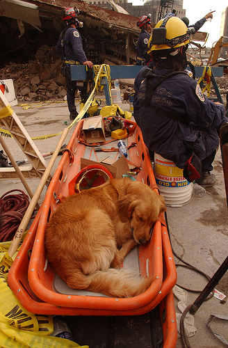 Four-Legged Heroes: Dangers for Search-and-Rescue Dogs
This article is courtesy of Mesothelioma.com.
Ofte
Four-Legged Heroes: Dangers for Search-and-Rescue Dogs
This article is courtesy of Mesothelioma.com.
Ofte
 Skin Problems in Dogs
By T. J. Dunn, Jr., DVM
What You Should Kno
Skin Problems in Dogs
By T. J. Dunn, Jr., DVM
What You Should Kno
Copyright © 2005-2016 Pet Information All Rights Reserved
Contact us: www162date@outlook.com