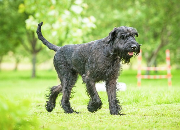
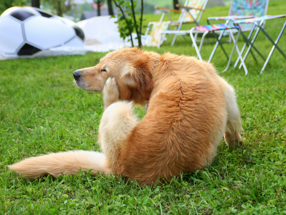
Cervical spondylomyelopathy (CSM), or wobbler syndrome, is a disease of the cervical spine (at the neck) that is commonly seen in large and giant-breed dogs. CSM is characterized by compression of the spinal cord and/or nerve roots, which leads to neurological signs and/or neck pain. The term wobbler syndrome is used to describe the characteristic wobbly gait (walk) that affected dogs have.
Intervertebral disk slippage and/or bony malformation in a narrowed vertebral canal (the bony canal surrounding the soft spinal cord) can cause spinal compression. Disk associated spinal compression is most often seen in dogs older than three years of age.
Doberman pinschers are predisposed to slipping intervertebral disks (in between the vertebrae). Vertebral malformation (bony associated compression) is most commonly seen in giant breed dogs, usually in young adult dogs that are less than three years of age. The bony malformation can compress the spinal cord from the top and bottom, from the top and sides, or just from the sides. Dynamic spinal cord compression (compression that changes with different positions of the cervical spine) always occurs with any type of compression.
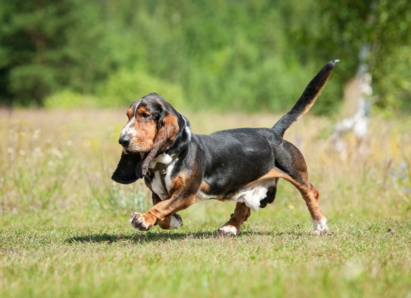
Breeds that appear to be predisposed to this condition are Doberman pinchers, rottweilers, great Danes, Irish wolfhounds, and basset hounds.
Along with the standard medical tests, which include a blood chemical profile, a complete blood count, a urinalysis and an electrolyte panel to rule out other diseases, your veterinarian will take a thorough history of your dog's health, onset of symptoms, and possible incidents that might have preceded this condition, such as traumas to the back or any previous illnesses. Any information you might have on your dog's genetic background may be helpful as well.
Wobbler syndrome is diagnosed via visualization. X-rays, myelographs, computed tomography (CT) and magnetic resonance imaging (MRI) will allow your doctor to view the spine and vertebrae. X-rays should be used mainly to rule out bony disorders while myelographs, CT and MRI are used to visualize the compression of the spinal cord. Diseases that will need to be ruled out though a differential diagnosis include diskospondylitis, neoplasia, and inflammatory spinal cord diseases. The results of the cerebral spinal fluid (CSF) analysis should pinpoint the origin of the symptoms.
Treatment will depend on the location of the spinal compression and the severity of the problem. If surgical treatment is not elected for, treatment may be given on an outpatient basis. Dogs that cannot walk should be kept on soft bedding, and should be closely observed and turned to lie on their other sides every four hours to prevent bed sores from developing.
Bladder catheterization may be used to allows the dogs to rest and not have to go outside to urinate. Your doctor will instruct you in how to do this procedure properly, with an emphasis on sterility to prevent urinary infections. Medically treated dogs typically need to have their activity restricted for at least two months. Surgery often offers the best chance of improvement (80 percent), but there is a small risk of significant complications associated with cervical surgical procedures.
Dogs which have had surgery should have their activity restricted two to three months postoperatively to allow bone ankylosis (adhesion and union) at the site of surgery. Physical therapy is essential for post-operative dogs to avoid muscle loss, atrophy, fusion of bones, and to hasten recovery. Your doctor will set up therapy sessions for your dog within the clinic, or will instruct you in methods by which you can help to maintain your dog's muscle integrity.
To protect your dog from further injury, do not allow any jumping or running for at least two to three months after after treatment. Body harnesses should be used in place of neck collars, since neck collars can harm your dog's already compressed spinal structure. Diet may also need to be adjusted. Cutting back on protein, calcium and excess calories is often recommended in dogs that are affected by CSM.
Your veterinarian will schedule follow up neurological evaluations as needed for your pet. If the symptoms of wobbler syndrome return, call your veterinarian immediately to be advised.
 Red Eye in Dogs
Inflammation of the Eye in Dogs
Red eye causes th
Red Eye in Dogs
Inflammation of the Eye in Dogs
Red eye causes th
 Heart Defect (Congenital) in Dogs
Patent Ductus Arteriosus in Dogs
The aorta is the
Heart Defect (Congenital) in Dogs
Patent Ductus Arteriosus in Dogs
The aorta is the
 Tick Paralysis in Dogs
Tick Bite Paralysis in Dogs
Ticks act as carriers
Tick Paralysis in Dogs
Tick Bite Paralysis in Dogs
Ticks act as carriers
 Parasite Drug (Ivermectin) Poisoning in Dogs
Ivermectin Toxicity in Dogs
This toxic reaction o
Parasite Drug (Ivermectin) Poisoning in Dogs
Ivermectin Toxicity in Dogs
This toxic reaction o
 Intestinal Parasite (Coccidia) in Dogs
Coccidiosis in Dogs
Coccidiosis is a parasitic ty
Intestinal Parasite (Coccidia) in Dogs
Coccidiosis in Dogs
Coccidiosis is a parasitic ty
Copyright © 2005-2016 Pet Information All Rights Reserved
Contact us: www162date@outlook.com