The Leydig cell tumor (LCT) is a rare, and typically benign (non-spreading) tumor made up from the cells that release the testosterone hormone in the connective tissue of the testicles. This type of tumor may occur singularly, or in multiples. The tumor mass is located in the testis, causing soft swelling of the affected testis. It measures about 1-2 cm in diameter and is spherical in shape. It is classified as a sex-cord stromal tumor, meaning that the tumor issues from the connective tissues of the sex-cords of the testis. This condition affects older male dogs.
The cause of LCT is unknown, but a retained testicle (usually in the abdomen) may predispose an animal to Leydig cell tumor formation.
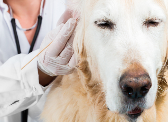
Your veterinarian will perform a complete physical exam, palpating (examination by touch) your dog’s testicles to examine the size, location, and consistency of the tumor. You will need to provide a thorough history of your dog’s health, with a description of symptoms, if any, and their onset. Usually a blood chemical profile, complete blood count, and urinalysis will return as normal, but there may be various reductions of cells in the circulating blood if there is an excess of estrogen. Blood serum should also be tested for estradiol, an estrogenic hormone, and testosterone concentrations. Usually estradiol levels will be high, while testosterone levels will be low. Your veterinarian may also take a fine needle sample of fluid (aspirate) from the tumor to check for abnormalities in the cells, by use of a cytological (microscopic) examination
Tumors smaller than 3 cm in diameter will appear black on ultrasound imaging. However, tumors greater than 5 cm have a black and white blotched appearance on ultrasound.
Affected dogs should be neutered, and the tumor(s) removed and sent for histopathologic analysis – examination of the diseased tissue. If your dog shows signs of bone marrow underdevelopment, your veterinarian may prescribe medical therapy to reverse it.
You will need to observe the post-operative surgical incisions for swelling, redness or oozing. Infection after surgery is always a cause for concern. If any of these conditions are present, or if you have any questions, contact your veterinarian for further advisement. If you are concerned that you may not be able to prevent your dog from getting its surgical site dirty, cage rest is an option. In this way you can be sure that your dog is staying clean, and is resting in an enclosed environement while the surgery wound heals. Additionally, you may want to use an Elizabethan collar to prevent your dog from being able to scratch or bite at the skin as it heals.
 Bacterial Infection (Mycoplasma, Ureaplasma, Acoleplasma) in Dogs
Mycoplasmosis in Dogs
Mycoplasmosis is the genera
Bacterial Infection (Mycoplasma, Ureaplasma, Acoleplasma) in Dogs
Mycoplasmosis in Dogs
Mycoplasmosis is the genera
 Coughing in Dogs
Tussis in Dogs
The act of coughing serves as a pr
Coughing in Dogs
Tussis in Dogs
The act of coughing serves as a pr
 Carbon Monoxide Poisoning in Dogs
Carbon Monoxide Toxicosis in Dogs
Carbon monoxide
Carbon Monoxide Poisoning in Dogs
Carbon Monoxide Toxicosis in Dogs
Carbon monoxide
 Anemia Due to Chronic Kidney Disease in Dogs
Erythropoietin (EPO) is a glycoprotein hormone, p
Anemia Due to Chronic Kidney Disease in Dogs
Erythropoietin (EPO) is a glycoprotein hormone, p
 Leukemia (Acute) in Dogs
Acute Lymphoblastic Leukemia in Dogs
Acute lympho
Leukemia (Acute) in Dogs
Acute Lymphoblastic Leukemia in Dogs
Acute lympho
Copyright © 2005-2016 Pet Information All Rights Reserved
Contact us: www162date@outlook.com