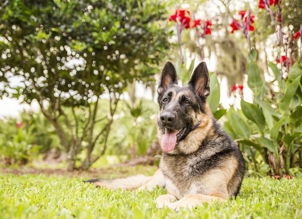
A histiocytoma is a benign skin tumor that originates in the Langerhans cells, immune cells that function to provide protective immunity to the tissues that are in contact with the outer environment -- the nose, stomach, intestines and lungs, but mainly the skin's surface. These cells are also referred to as dendritic cells, and histiocytes.
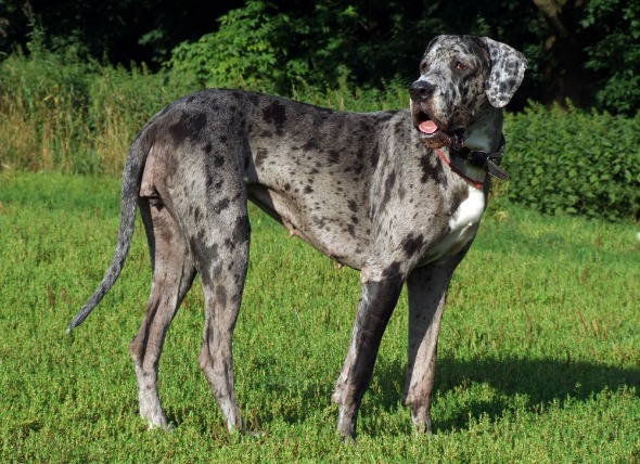
Histiocytomas are common in dogs, with some breeds appearing to be more predisposed that others. These breeds include flat-coated retrievers, bull terriers, boxers, dachshunds, cocker spaniels, Great Danes, and Shetland sheepdogs. More than 50 percent of diagnosed patients are under two years of age. Otherwise, there is no gender difference.
Unknown
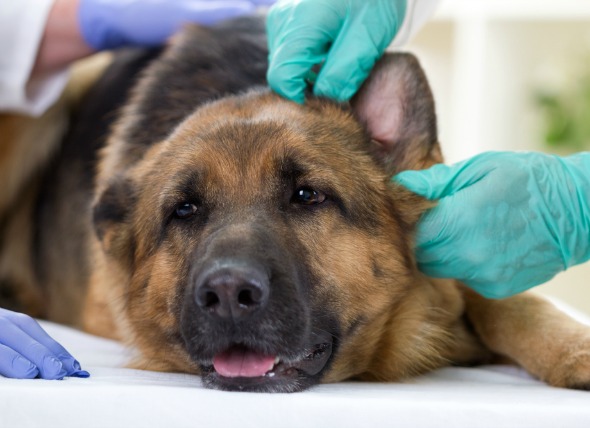
You will need to give a thorough history of your dog's health and onset of symptoms, after which your veterinarian will perform a thorough physical exam. A complete blood profile will be conducted, including a chemical blood profile, a complete blood count, a urinalysis and an electrolyte panel. Most of these tests return as normal.
Other diagnostic tests include a cytologic examination (a microscopic examination of the cells) using a sample gathered by fine-needle aspirate. This may reveal pleomorphic round cells (cells taking one or more forms), with variable-sized and -shaped nuclei. It is common to find that the mitotic index (a measure of the proliferation, or fast production status of a cell population) is high. The tests may also show evidence of substantial lymphocyte (white blood cell in the vertebrate immune system), plasma cell, and neutrophil (the most abundant type of white blood cells) infiltration.
Since some treatments can adversely affect malignant tumors, important to differentiate histiocytoma, a benign growth of tissue, from a malignant tumor. Your veterinarian will talk to you about this, and will give you the option of taking a wait-and-see approach. If you do have the tumor diagnosed conclusively, and it is found to be a histiocytoma, the usual method of treatment is surgical excision of the mass, or cryosurgery, which is conducted with a laser. Either one is generally curative.
If the mass is left alone, it may spontaneously regress within three months. This is a decision that you will have to make once you have been informed of every possible eventuality, and every treatment method that is available for your dog.
Surgical excision of the mass is recommended if the mass has not spontaneously regressed within three months. With removal of the mass, the long-term prognosis is generally excellent.
 Inflammatory Skin Disease in Dogs
Sebaceous Adenitis in Dogs
Sebaceous adenitis is
Inflammatory Skin Disease in Dogs
Sebaceous Adenitis in Dogs
Sebaceous adenitis is
 Blood Transfusion Reactions in Dogs
There are a variety of reactions that can occur w
Blood Transfusion Reactions in Dogs
There are a variety of reactions that can occur w
 Long-Term Stomach Inflammation in Dogs
Chronic Gastritis in Dogs
Chronic gastritis is th
Long-Term Stomach Inflammation in Dogs
Chronic Gastritis in Dogs
Chronic gastritis is th
 High Blood Pressure in Dogs
Systemic Hypertension in Dogs
More commonly refer
High Blood Pressure in Dogs
Systemic Hypertension in Dogs
More commonly refer
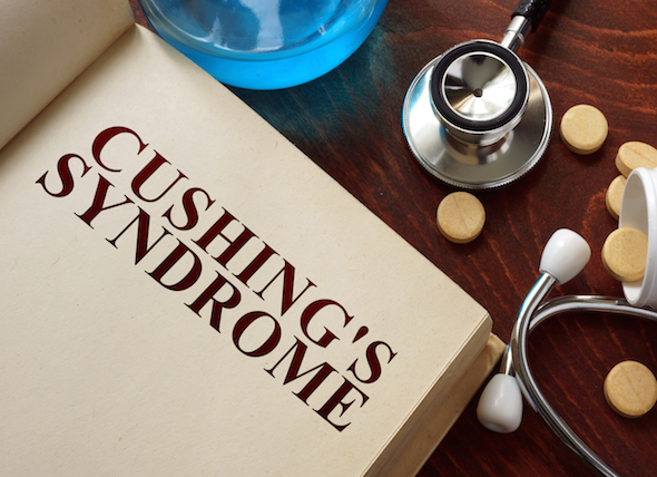 Cushing's Disease in Dogs
Hyperadrenocorticism in Dogs
The endocrine system
Cushing's Disease in Dogs
Hyperadrenocorticism in Dogs
The endocrine system
Copyright © 2005-2016 Pet Information All Rights Reserved
Contact us: www162date@outlook.com