Cyanosis is a medical condition characterized by blue colored skin and mucous membranes, which occurs as the result of inadequate amounts of oxygenated hemoglobin -- the molecule which carries oxygen to the body tissues -- or due to hemoglobin abnormalities.
Unfortunately, dogs that are suffering from cyanosis caused by advanced lung/airway disease and severe heart disease have a poor long-term prognosis.
Originating in the Respiratory System
Originating in the Cardiovascular System
Originating in the Neuromusculoskeletal System
Methemoglobinemia
Your veterinarian will first stabilize your dog's oxygen levels. This is usually done in ICU (intensive care unit) in a specially equipped oxygen cage. Once your dog is stable, your veterinarian will be able to perform a full physical exam.
A blood chemical profile, complete blood count, urinalysis, electrocardiograph (EKG), thoracic radiographs (and echocardiogram with Doppler, if heart or lung disease is suspected), and an electrolyte panel should be ordered to determine the underlying cause of the disease that is causing cyanosis.
A laryngoscopic (voice box) and/or bronchoscopic (lung airway) exam should be given. If bronchopulmonary (lung disease) disease is suspected, a transtracheal wash, a bronchoalveolar lavage or fine-needle lung aspirate may be performed. For pleural space disorders, a thoracocentesis (a procedure which removes fluid from the chest cavity) will be required.
Methemoglobinemia is a condition that can be measured; one of the most obvious indications is that the color of the blood will be darker than the bright red it is supposed to be. Arterial blood can be taken so that a blood gas analysis can be performed at the laboratory. Your dog's breathing patterns will also give your veterinarian a clue as to the origin of the cyanosis.
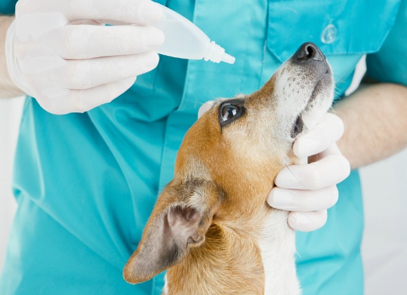
Your dog will need to be kept stabilized by giving it oxygen. Depending on what underlying illness is causing the cyanosis, drugs may be prescribed to treat the condition, or surgery and/or further therapy ordered.
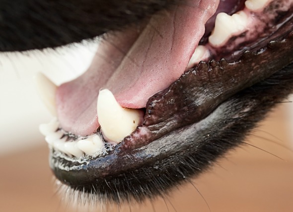
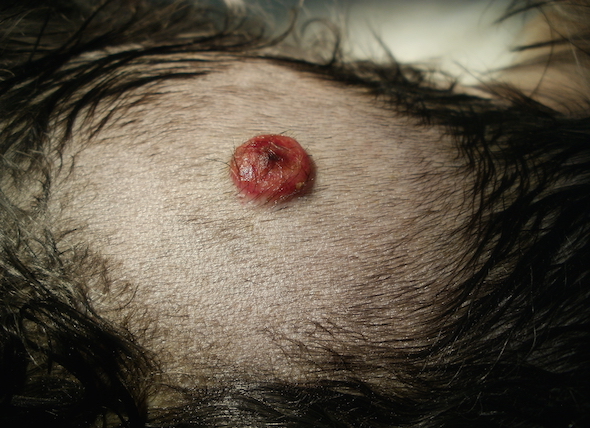
You will need to restrict your dog's activity during treatment and possibly after. A low-salt diet can be put in place if your veterinarian determines that heart disease is involved. You should also check your dog's gums for normal color, making sure they are a healthy pink or reddish color. If your dog's gums are purple or white, you should take it immediately to the veterinary hospital for emergency treatment.
 Hemorrhage of the Lung in Dogs
Pulmonary Contusions in Dogs
Pulmonary contusion,
Hemorrhage of the Lung in Dogs
Pulmonary Contusions in Dogs
Pulmonary contusion,
 Acute (Sudden) Dog Diarrhea
Dog Diarrhea has four general reasons for occurring: osm
Acute (Sudden) Dog Diarrhea
Dog Diarrhea has four general reasons for occurring: osm
 Seizures and Convulsions in Dogs
Status Epilepticus in Dogs
Status epilepticus, or
Seizures and Convulsions in Dogs
Status Epilepticus in Dogs
Status epilepticus, or
 Coonhound Paralysis In Dogs
Idiopathic Polyradiculoneuritis in Dogs
Acute can
Coonhound Paralysis In Dogs
Idiopathic Polyradiculoneuritis in Dogs
Acute can
 Brain Injury in Dogs
Dogs can incur brain injuries from a variety of c
Brain Injury in Dogs
Dogs can incur brain injuries from a variety of c
Copyright © 2005-2016 Pet Information All Rights Reserved
Contact us: www162date@outlook.com