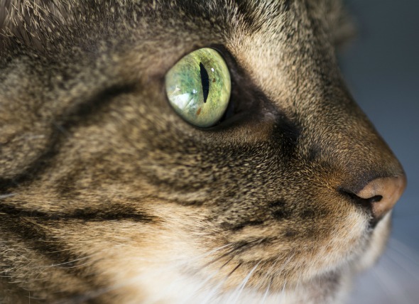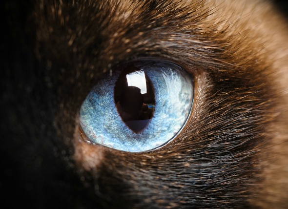
The uvea is the part of the eye that is made up of the iris (the colored part of the eye surrounding the pupil), the ciliary body (which produces the fluid within the eye [aqueous humour] and controls the ciliary muscle contractions that aid in near focus), the choroid (which provides oxygen and nourishment to the retina – the inner surface of the eye), and the pars plana (at the front of the eye, where the iris and sclera [white of the eye] touch). A melanoma is clinically characterized by malignant growth of melanocytes, cells that are dark in appearance due to the inclusion of the melanin pigment.
Uveal melanomas in cats usually arise from the front of the iris’ surface, with extension to the ciliary body and choroid. These tumors tend to be flat and diffuse, not nodular (unlike intraocular melanomas, which are raised masses). Such tumors initially have a benign (non-spreading) clinical and cellular appearance. However, an affected cat may develop metastatic disease (due to the spread of the uveal melanoma) up to several years later. The metastatic rate may be up to 63 percent. These tumors are also called diffuse iris melanomas – that is, melanomas of the iris that are capable of spreading. This is the most common type of eye tumor in cats.
Your veterinarian will perform a thorough physical exam on your cat, including a complete ophthalmic exam (including testing pressure within the eye and proper drainage of the eye’s aqueous humor). During the ophthalmic exam, tonometry will be used to measure the pressure in the eyes, and gonioscopy will be used to see if the melanoma has spread to the drainage angle. Slit-lamp biomicroscopy can be used to gage the size and location of the mass.
A complete blood profile will also be conducted, including a chemical blood profile, a complete blood count, a urinalysis and an electrolyte panel. Evidence of metastasis may may be present in the blood profile, or the blood count may show increased white blood cells, which can be indicative of the body's immune system fighting the malignant cell growth. You will need to give a thorough history of your cat's health and onset of symptoms.
X-rays and an ultrasound may also help to determine the extent of metastatic disease in the eye. If there are melanoma cells in the angle between the iris and the cornea, and if there are melanoma cells in the ciliary venous plexus (where veins from the ciliary body drain blood from the eye), then metastatic (cancerous) cells have probably spread throughout the body. However, this metastasis may not be evident until a few years after the initial growth of cells.
One long-term study shows that patients with early iris melanoma have no increased risk of life-threatening cancer spread compared to controls, but patients with advanced lesions had dramatically shortened survival times. Your cat's prognosis will depend on if and how much the melanoma has spread within the eye. Your veterinarian will schedule follow-up appointments with you every three months to monitor your cat's intraocular pressure if you decline to have surgery performed on the eye. X-rays to check for metastasis should be taken every six months after the initial diagnosis was made.


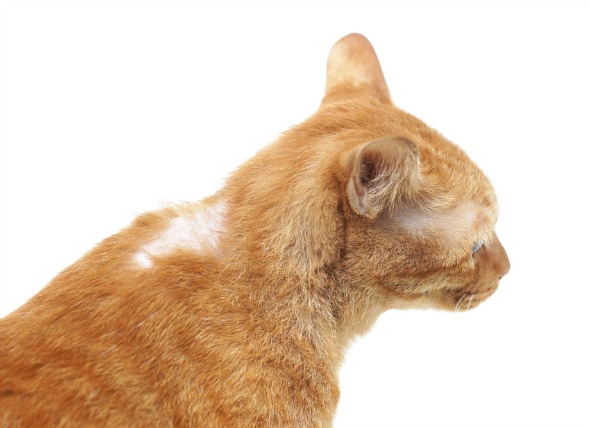 Hair Loss Related to Cancer in Cats
Feline Paraneoplastic Alopecia in Cats
Feline par
Hair Loss Related to Cancer in Cats
Feline Paraneoplastic Alopecia in Cats
Feline par
 Bone Cancer (Chondrosarcoma) in Cats
Chondrosarcoma of the Bone in Cats
Chondrosarcoma
Bone Cancer (Chondrosarcoma) in Cats
Chondrosarcoma of the Bone in Cats
Chondrosarcoma
 Teeth Misalignment in Cats
Malocclusion of Teeth in Cats
Normally, a kitten
Teeth Misalignment in Cats
Malocclusion of Teeth in Cats
Normally, a kitten
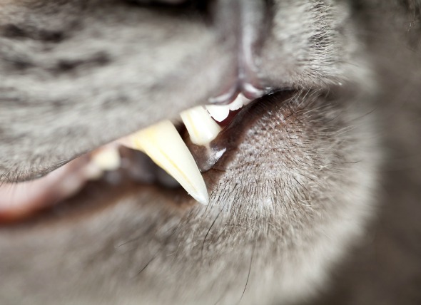 Tooth Decay in Cats
Feline Oral Resorptive Lesions (FORL) or Odontocl
Tooth Decay in Cats
Feline Oral Resorptive Lesions (FORL) or Odontocl
 Rabies in Cats
Rabies is an inflammatory infection that specific
Rabies in Cats
Rabies is an inflammatory infection that specific
Copyright © 2005-2016 Pet Information All Rights Reserved
Contact us: www162date@outlook.com