Ear hematomas, also known as auricular hematomas or aural hematomas, occur when blood accumulates in the flap (or pinna) of the ear.
Characterized by a swelling of the ear flap, ear hematomas often occur in only one ear. However, it is possible for both ears to have hematomas. The swelling may involve the entire ear flap or it may cover only part of the ear flap.
The most common cause of an ear hematoma in cats is an ear mite infection. Ear mites cause irritation to the ear, resulting in shaking of the head which in turn causes the development of the hematoma. Other ear infections can also be responsible for hematoma formation.
Less commonly, allergic skin disease, immune disorders or blood clotting deficits can be the cause of ear hematomas.
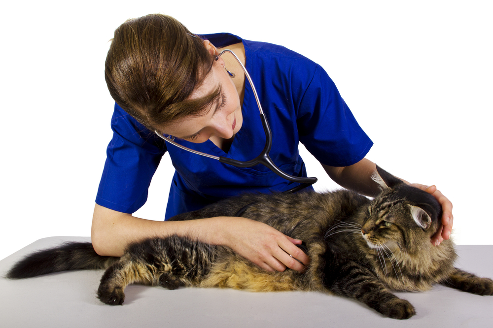
Ear hematomas are usually easily diagnosed by physical examination.
Many different treatments exist for ear hematomas. The fluid within the hematoma can be drained but the hematoma is likely to recur and may need to be drained numerous times. Many veterinarians prefer to lance the hematoma and drain the fluid under anesthesia. In most cases, a drain is placed in the ear to keep additional fluid from building up within the ear flap or, alternatively, sutures or other devices may be placed through the ear flap to discourage additional accumulation of fluid and recurrence of the hematoma.
If ear disease is present, it will need to be treated simultaneously.
Preventing ear infections is often effective in preventing ear hematoma formation. When ear infections do occur, they should be treated promptly to avoid the formation of a hematoma.


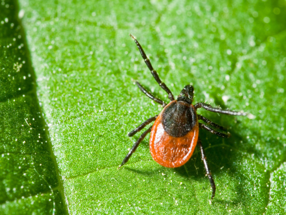 Ticks and Tick Control in Cats
Ticks are parasitic organisms that attach themsel
Ticks and Tick Control in Cats
Ticks are parasitic organisms that attach themsel
 Painful Abdomen in Cats
Peritonitis in Cats
Acute pain in the abdomen due
Painful Abdomen in Cats
Peritonitis in Cats
Acute pain in the abdomen due
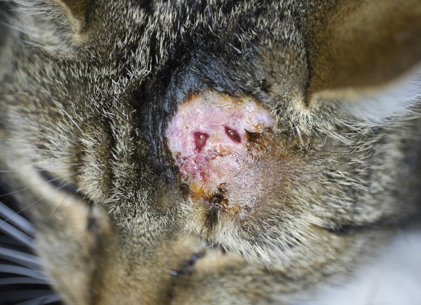 Abscesses in Cats
Cats, like people, are prone to skin irritations.
Abscesses in Cats
Cats, like people, are prone to skin irritations.
 Why Do Cats Spray Indoors?
Why Do Cats
Why Do Cats Spray Indoors?
Why Do Cats Spray Indoors?
Why Do Cats
 Aggression in Cats (Overview)
Cats are small, and often become the target of ot
Aggression in Cats (Overview)
Cats are small, and often become the target of ot
Copyright © 2005-2016 Pet Information All Rights Reserved
Contact us: www162date@outlook.com