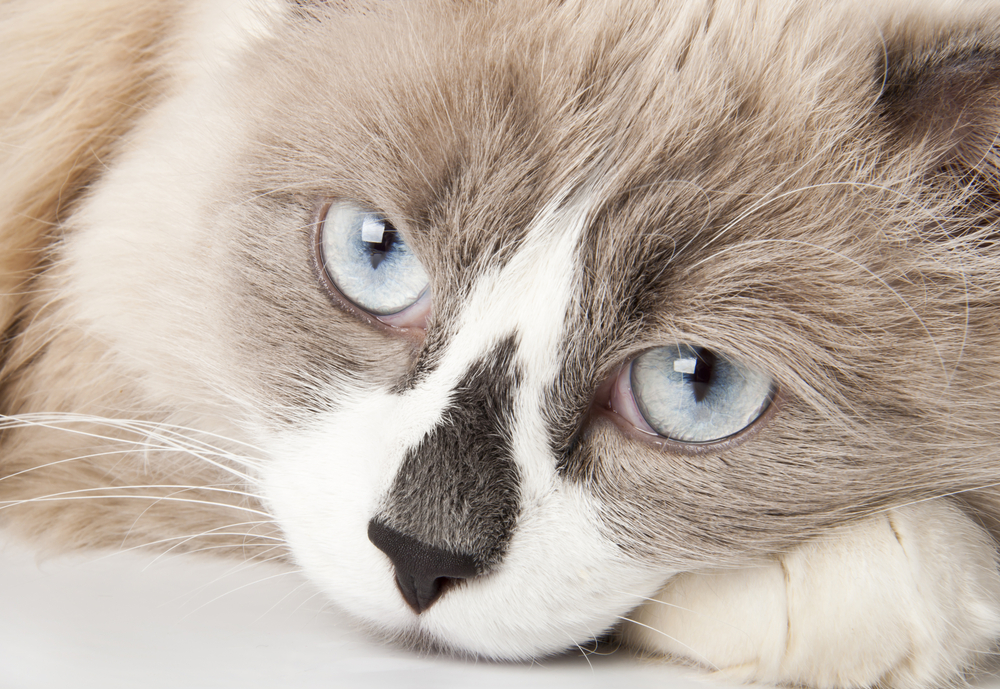
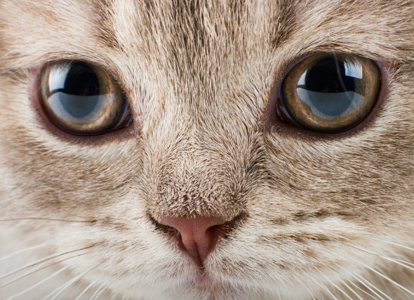
Keratitis is the medical term given to inflammation of the cornea -- the clear outer layer of the front of the eye. Nonulcerative keratitis is any inflammation of the cornea that does not retain fluorescein stain, a dye that is used to identify ulcers of the cornea. If the very top layer of the cornea has been disrupted (as with an ulcer), the dye will enter the lower layers of the cornea and will cause a temporary stain that glows under an ultraviolet light; in nonulcerative keratitis, the top layer of the cornea is not disrupted, so no dye enters the lower layers of the cornea.
Long-term superficial inflammation of the cornea may occur at any age, but the risk is higher between the ages of four to seven years. There are different forms that nonulcerative keratitis can take. Inflammation involving the area where the cornea (clear part of the eye) and the sclera (white part of the eye) come together, and characterized by the presence of nodules, may occur. Another is a condition in which part of the corneal tissue dies, leaving a pigmented lesion and fluid build-up. These forms can occur at any age, but the latter form is most prevalent in Persian, Siamese, Burmese, and Himalayan breeds. There is no proven genetic basis in cats that has been found so far. However, geographical location has been found to play some role, as animals living at higher altitudes appear to be at increased risk.
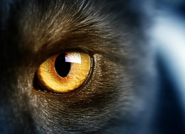
Herpesvirus in cats can affect cats of all ages and can lead to inflammation of the cornea. This form is characterized by the presence of a type of white-blood cell called an eosinophil (condition known as eosinophilic keratitis) and results in a condition in which part of the corneal tissue dies, leaving a pigmented lesion and fluid build-up in the eye. This can occur at all ages except in newborns.
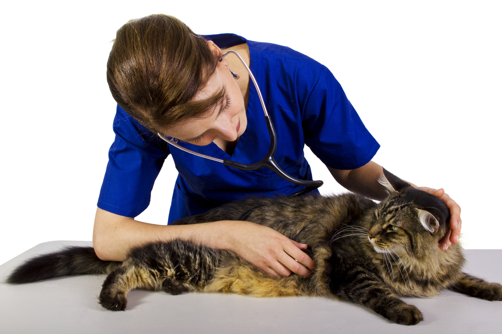
Your veterinarian will perform a thorough physical and ophthalmological exam on your cat, taking into account the background history of symptoms and possible incidents that might have led to this condition. Cultures of cells will be conducted to determine whether there are excess white blood cells (indicating a physical response to an invasive condition) or organisms present in the bloodstream. A biopsy of the cornea may also be done, although your veterinarian will probably be able to make a diagnosis without it.
Your cat will only need to be hospitalized if it does not respond adequately to medical therapy. Outpatient care is generally sufficient. Radiation therapy may be prescribed for long-term superficial inflammation of the cornea. Radiation therapy and cryotherapy (a freezing technique that is used for removal of diseased tissue) may also be prescribed for inflammation characterized by the presence of pigment that is deposited in the cornea.
If the diagnosis is inflammation of the cornea characterized by the presence of a type of white-blood cell called an eosinophil, surgical removal of the surface of the cornea may be done for diagnostic purposes. This is usually unnecessary as it only temporarily resolves clinical signs; medical treatment is preferred.
If the condition takes the form in which part of the cornea tissue is dying, leaving a pigmented lesion and fluid build-up, surgical removal of the surface of the cornea may be curative, but recurrence is possible; eye discomfort is the primary indication for surgery.
There are medications that your veterinarian may prescribe as a part of the treatment regimen for the various forms of this condition, depending on what the final diagnosis is.
Long-term superficial inflammation of the cornea is more likely to occur at high altitudes with intense sunlight.
Your veterinarian will want to conduct periodic eye examinations to evaluate the effectiveness of treatment. Your doctor will set up a follow-up schedule to see your cat at one to two week intervals, gradually lengthening the interval as long as your cat remains in remission, or the clinical signs resolve. In severe cases your cat may have continued eye discomfort, some visual defects, and in some cases, may even suffer from permanent blindness.
 Why Cats Scream
Why Cats Scream. Cats,
Why Cats Scream
Why Cats Scream
Why Cats Scream. Cats,
 Colonic or Rectal Inflammation in Cats
Colitis and Proctitis in Cats
Histiocytic ulcerat
Colonic or Rectal Inflammation in Cats
Colitis and Proctitis in Cats
Histiocytic ulcerat
 Lily Plant Poisoning in Cats
Toxic Reaction to Lily House Plants
One of the mo
Lily Plant Poisoning in Cats
Toxic Reaction to Lily House Plants
One of the mo
 What Kind Of Human Food Can Cats Eat?
People Food For CatsMany cat
What Kind Of Human Food Can Cats Eat?
People Food For CatsMany cat
 Chest Bone Deformity in Cats
Pectus Excavatum in Cats
The sternum, or chest bo
Chest Bone Deformity in Cats
Pectus Excavatum in Cats
The sternum, or chest bo
Copyright © 2005-2016 Pet Information All Rights Reserved
Contact us: www162date@outlook.com